Page 236 - 2021_09-Haematologica-web
P. 236
Letters to the Editor
A
B
CD
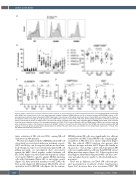
Figure 2. CD56+ natural killer cells from multiple myeloma patients are hypo-responsive to elotuzumab-labeled myeloma cells. Peripheral blood mononuclear
cells (PBMC) from healthy donors (n=9), and newly diagnosed mulptiple myeloma (NDMM) patients (n=10) or refractory relapsed MM (RRMM) patients (n=10) at baseline (pre-treatment) were cultured with OPM2 target cells in the presence of 10 μg/mL elotuzumab (elo) or human IgG1 (iso) isotype control. Shown in (A) histogram overlay of changes in CD16 expression on CD56dimCD16+ subset of natural killer (NK) cells. (B) Percentage distribution of NK-cell subsets (left panel) or percentage of CD16+ on CD56dimCD57+ NK cells (right panel) in healthy donor (HD), newly diagnosed MM and RRMM patient PBMC after treatment under the same conditions as above. (C) Percentage distribution of CD56dimCD16+ subset of NK cells in HD and RRMM patient PBMC after treatment under the same conditions as above in the presence or absence of ADAM17 inhibitor (n=5 per group) (D) Collated data for HD, NDMM and refractory relapsed (RR) MM patients (n=9-10 per group) showing CD107a degranulation by different NK-cell subsets. Data are pooled from four independent experiments. *P<0.05, One- way ANOVA with Bonferroni post-hoc test.
tutive activation of NK cells via CD16, causing NK-cell exhaustion in MM patients.
We then investigated whether NDMM patient NK-cell cytotoxicity recovered post-induction treatment or post- ASCT and if they can be targeted with monoclonal anti- body therapy. In order to reveal myeloma patient NK cell killing potential, we investigated their cytotoxicity against the MHC class I negative erythro-leukemia cell line, K562 (Figure 3A), their antibody-dependent cellular cytotoxicity (ADCC) capacity against OPM2 myeloma cells with elotuzumab (Figure 3B), or an isotype control (Figure 3C). After induction treatment or ASCT, NK cells from newly diagnosed MM patients killed K562 cells at equivalent levels to HD NK cells (Figure 3A). In contrast,
NDMM patient NK cells were significantly less efficient at myeloma cell ADCC than HD NK cells, requiring high- er numbers of NK effectors to achieve target lysis (Figure 3B); this reduced ADCC function was present after induction therapy, and after ASCT (Figure 3B). Finally, in the presence of an isotype control, myeloma patient NK cells were significantly less efficient than HD NK cells at killing myeloma targets (Figure 3C). Taken together, this data suggests whilst myeloma patient NK cells have cyto- toxic potential, they are unable to effectively kill myelo- ma targets.
There was no difference in CD16+ NK cells from pre- and post-induction treatment (Online Supplementary Figure S3C); however, there were less mature CD57+ NK
2524
haematologica | 2021; 106(9)


