
Page 220 - 2021_09-Haematologica-web
P. 220
Letters to the Editor
A
B
CD
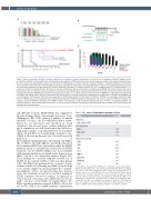
Figure 2. Effects of ruxolitinib, S63845, and their combination on inhibition of plasma cell growth in vitro and in vivo. (A) Inhibition of INA-6.Tu1 growth in vitro by ruxolitinib, S63845, or their combination. Cell growth was measured by an MTS-based colorimetric assay. Drugs were added at the indicated concentrations. The mean values of a representative experiment, performed in quadruplicate, are shown. Error bars, standard deviation. *Significant difference of the effect of the combination from the effect of either single drug (P<0.05, unpaired two-tailed t-test). The drug combination indices (CI) for experimental values at a con- stant ratio were calculated with the method of Chou and Talalay with CalcuSyn v2.0 software (Biosoft, UK). CI<1, synergistic effect; CI=1, additive effect; CI>1, antagonistic effect. Fa: affected fraction. (B) Treatment scheme of SCID mice with the combination of ruxolitinib and S63845. Treatment started at day 1 after intraperitoneal (IP) cell inoculation and continued for 10 consecutive days. Ruxolitinib was administered orally (PO) twice daily, with the time between two doses being approximately 6 h. S63845 was injected intravenously (IV) on days 1, 4, 7, and 10. Treatment with ruxolitinib or S63845 as single agents was performed accordingly with vehicle always used as a substitute for the second drug. (C) Survival of SCID mice treated with the combination of ruxolitinib and S63845 (red line) was superior (100% alive) to that of animals treated with ruxolitinib alone (green line; 43% alive; P=0.0325) or S63845 as a single agent (purple line; 50% alive; P=0.0514). The control group (black line) received vehicle (0% alive; P≤0.0001 against all other groups). There was no significant difference between the ruxolitinib- and the S63845-treated group (P=0.4768). P-values were calculated using the log-rank (Mantel-Cox) test: P<0.05 is considered significant. (D) Soluble interleukin-6 (IL-6) receptor levels in the serum of mice at the day of sacrifice. Animals with undetectable levels had no visible tumors and survived until the experiment was terminated.
salt (INC424; formerly INCB018424) was supplied by Novartis Pharma (Basel, Switzerland) and Incyte Corp. (Wilmington, DE, USA). Among a number of human myeloma cell lines, the IL-6-dependent INA-6 (estab- lished in our laboratory and described in detail elsewhere5) was chosen because cytokine pathways after gp130 stimulation are well characterized and the line is sufficiently sensitive to growth inhibition by ruxolitinib (Figure 1A and Table 1). A similar high sensitivity to rux- olitinib in the nanomolar range was observed for growth inhibition of IL-6-stimulated primary plasma cell leukemia cells (Figure 1B). In INA-6, the JAK inhibitor specifically abrogated IL-6-stimulated STAT3 phosphorylation while the MAPK pathway, which is constitutively activated by an N-RAS mutation,5 was not inhibited (Figure 1C). Concomitantly with signaling inhibition, ruxolitinib induced apoptosis in INA-6 cells in a dose-dependent manner (Figure 1D). These findings are consistent with the essential role of STAT3 for the survival of INA-6 cells6 and other plasma cells.7 The INA-6 xenograft model also seemed to be par- ticularly suitable for evaluating ruxolitinib given the high in vivo activity of gp130 monoclonal antibodies.8 As phar- macodynamic studies on tumor-bearing mice demon- strate, the constitutive as well as (ex vivo) IL-6-stimulated STAT3 activation observed in tumors of untreated or vehicle-treated control mice were inhibited in vivo by one single oral dose of ruxolitinib (60 mg/kg) (Figure 1E). Other signaling pathways activated in INA-6 cells in vitro and in vivo, such as the MAPK pathway constitutively
Table 1. IC50 values of ruxolitinib in myeloma cell lines.
Cell line
JAK driven
HEL (JAK2 V617F)
IL-6 dependent INA-6
INA-6.Tu1
B9
Autonomous growth
EJM
JJN3
JK-6
L363 MM1.S NCI-H929 RPMI8226 U266
IC50 (μM)
0.8
0.15 0.85 0.6
2.67 >8* 4.19 >8* >8* >8* >8* >8*
Cell growth was measured by an MTS-based colorimetric assay and half maximal inhibitory concentration (IC50) values were calculated with CalcuSyn v2.0 software (Biosoft, UK). The erythroleukemia line HEL carrying the activating JAK2 V617F mutation served as a control.With the exception of the murine B9 hybridoma,all cell lines were of human origin. B9 was a kind gift from L. A. Aarden (Central Laboratory Blood Transfusion Service, Amsterdam, the Netherlands); MM1.S was kindly provided by Yu Tzu Tai (Dana Farber Cancer Institute, Boston, MA, USA); INA- 6, INA-6.Tu1 and JK-6 were established as described elsewhere.5,16 All other cell lines were obtained from the German Collection of Microorganisms and Cell Cultures (DSMZ), Braunschweig, Germany. *Highest concentration evaluated.
2508
haematologica | 2021; 106(9)
