
Page 219 - 2021_09-Haematologica-web
P. 219
Letters to the Editor
Dual intracellular targeting by ruxolitinib and the Mcl-1 inhibitor S63845 in interleukin-6-dependent myeloma cells blocks in vivo tumor growth
Multiple myeloma remains an incurable malignancy with most patients experiencing relapse despite the intro- duction of novel therapies. While the first monoclonal antibodies have been approved for the treatment of myeloma, small molecule inhibitors of signaling path- ways are still investigational. Although the concept of Janus kinase (JAK)/signal transducer and activator of transcription (STAT)3 inhibition in myeloma has shown promising results in preclinical studies, the efficacy of
JAK inhibitors as single agents seems to be limited.1 Ruxolitinib is a potent JAK1/2 inhibitor and approved for the treatment of patients with myeloproliferative disease and for graft-versus-host disease.2 While it has activity as a single agent in multiple myeloma, the combination with the myeloid cell leukemia (Mcl)-1 protein inhibitor S63845 resulted in superior survival in a preclinical in vivo model. The results obtained in the INA-6 xenograft model strongly support evaluation of the combination of JAK and Mcl-1 inhibition in humans.
The JAK/STAT3 pathway is activated by cytokines of the gp130 family including interleukin (IL)-6 as the most prominent member with an established pathophysiolog- ical role in multiple myeloma.3,4 Ruxolitinib phosphate
AB
CD
E
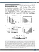
Figure 1. Effects of ruxolitinib on malignant plasma cell growth and STAT3 phosphorylation in vitro and in vivo. (A) Inhibition of INA-6 growth in vitro by ruxoli- tinib is dose-dependent. Cells were cultured in the presence of 2.5 ng/mL interleukin-6 (IL-6) for 3 days and absorbance was measured in an MTS-based col- orimetric assay as described elsewhere.8 The mean values of ten independent experiments, each performed in triplicate or quadruplicate, are shown. Error bars, standard deviation. The concentration at 50% inhibition was calculated with CalcuSyn software (Biosoft, UK). (B) Inhibitory effect of ruxolitinib on IL-6-stimulated proliferation of primary plasma cells from the peripheral blood of a patient with plasma cell leukemia. 3H-thymidine uptake was measured as described previ- ously.5 (C) Ruxolitinib dose-dependently inhibits IL-6-induced STAT3 phosphorylation in INA-6 cells, as demonstrated by western blot analysis. INA-6 cells were starved of IL-6 and serum for 4 hours (h), treated with different concentrations of ruxolitinib for 2 h, and then stimulated with 10 ng/mL IL-6 (Gibco®/Life Technologies, Darmstadt, Germany) for 15 min. Control cells did not receive IL-6. Cropped blots are shown. (D) Induction of apoptosis by ruxolitinib as shown by annexin V-FITC/7-AAD staining (Beckman-Coulter) and flow cytometric analysis (FC500). Cells were cultured in IL-6 and different concentrations of ruxolitinib for 48 h and 72 h. Control cells (Ctrl.) did not receive IL-6 or ruxolitinib. (E) Inhibition of STAT3 phosphorylation in vivo. A single oral dose of ruxolitinib (60 mg/kg) was given to tumor-bearing mice (at day 27 or day 33 after cell inoculation). One control animal received vehicle (0.5 % w/v methylcellulose, day 33), one engraft- ed mouse remained untreated. Tumors were explanted 2 h after drug administration. One part of the cells was stimulated ex vivo with IL-6 (10 ng/mL) for 10 min (+), the other part remained unstimulated (-). Cell lysates were prepared for sodium dodecylsulfate polyacrylamide gel electrophoresis and western blot analysis. Cropped blots of cell lysates from the two control animals and two ruxolitinib-treated mice are shown.
haematologica | 2021; 106(9)
2507
