
Page 195 - 2021_09-Haematologica-web
P. 195
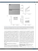
Activation of SCD RBC adhesion by oxidative stress
A
B
Figure 3. Effect of in vitro oxidation on AA red blood cell adhesion to laminin. (A) Left panel: typical microscopy images of non-oxidized and cumene hydroperoxide- oxidized AA red blood cells (RBC) adhering to Laminin 521 at 3 dyn/cm2. Right panel: quantification of cell adhesion showing the mean number of adherent RBC/mm2 in seven oxidized and non-oxidized AA RBC samples at 3 dyn/cm2. Wilcoxon test, *P<0.05. (B) Flow cytometry analysis of Lu/BCAM expression at the RBC surface expressed as percentage of Lu/BCAM-positive RBC (left panel) and mean fluorescence intensity (MFI) of these RBC (right panel) under non-oxidized (phosphate buffered saline [PBS]) and oxidized conditions (cumene). No significant differences were observed, Wilcoxon test, P=0.0714.
In order to gain insight into the potential mechanism underlying this difference between LD and HD RBC, cells were fixed after the 7 dyn/cm2 step, stained fluorescently for Lu/BCAM and analyzed by confocal microscopy. There was a difference in the expression pattern of Lu/BCAM between LD and HD RBC at the interface with laminin. HD RBC showed a homogeneous distribution of Lu/BCAM with some cells showing intense staining and the presence of bigger spots suggestive of potential Lu/BCAM aggregates (Figure 2E; Online Supplementary Figure S2B). LD RBC showed cells with a smaller surface contact area, large fluorescent patches and very fine fluo- rescent membrane extensions tethering the cells to the surface (Figure 2E). The proportion of RBC exhibiting membrane tethers was higher in LD (55.6%) than in HD RBC (8.2%) (Figure 2F), and tethers were also longer in LD RBC (Figure 2G) suggesting a more dynamic lipid bilayer in LD RBC.
Oxidation activates AA red blood cell adhesion to laminin
We have previously shown that Lu/BCAM-mediated adhesion to laminin is activated by phosphorylation of serine 621 of its cytoplasmic tail.17 In SCD, this phospho- rylation takes place in reticulocytes, with very low levels
of phosphorylation detected in HD RBC.27 Considering the high levels of adhesion of HD RBC, we hypothesized that Lu/BCAM might be activated by post-translational modifications triggered by oxidative stress. In order to test this hypothesis, we assessed adhesion of control (AA) RBC under oxidative conditions after incubation with cumene hydroperoxide (270 μM), an agent that induces membrane lipid peroxidation. AA RBC showed the expected residual adhesion to laminin, that was signifi- cantly increased after incubation with cumene hydroper- oxide (Figure 3A; Online Supplementary Figure S3). This was not due to a difference in the Lu/BCAM expression level between oxidative and control conditions as determined by flow cytometry measuring the percentage of Lu/BCAM-positive RBC and their mean fluorescence intensity (MFI) (Figure 3B). Moreover, this increased adhe- sion was not due to increased phosphorylation of Lu/BCAM as determined by western blot using an anti- phosphoSerine antibody (not shown).
Oxidative stress alters Lu/BCAM distribution at the red blood cell surface
Analysis of Lu/BCAM membrane distribution by confocal microscopy
We evaluated the impact of oxidation on the distribu-
haematologica | 2021; 106(9)
2483
