
Page 192 - 2021_09-Haematologica-web
P. 192
M.A. Lizarralde-Iragorri et al.
Confocal microscopy
Imaging was performed on the Confocal LSM 510 META-TIRF (Zeiss, Oberkochen, Germany). LASX software was used to set up and analyze the experiments (Leica microsystems, Wetzlar, Germany).
Imaging flow cytometry assays
Expression of Lu/BCAM on the RBC surface was analyzed using F241 mouse monoclonal antibody. After 1-hour incubation
with F241 (dilution [d]: 1/10), the secondary anti-mouse APC-con- jugated antibody (d: 1/100) (Beckman Coulter) was added for 1 hour, then RBC were washed and suspended in 200 μL of thiazole orange (TO) dye (Retic-CountTM, Becton-Dickinson) for 30 min- utes (min) to label reticulocytes. RBC were analyzed using ImageStream®X Mark II Imaging Flow Cytometer (Merck Millipore) (60x magnification) and the IDEAS software (version 6.2). Lu/BCAM-positive mature RBC (Lu APC) were gated, excluding the reticulocytes (TO-positive events). Using the
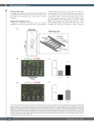
A
B
C
D
E
Figure 1. Analysis of red blood cell deformability using a microfluidic biomimetic chip. (A) Left panel: the microfluidic device comprises eight filtering units arranged in parallel. Right panel: each filtering unit is 5 μm-high and has a U shape composed of a series of 15 μm pillars separated by 5 μm slits, with two 10 μm-wide side channels. Inside the U shape, four rows are disposed in parallel with decreasing slit width (10, 8, 7 and 6 μm). (B) Microscopy image showing SS red blood cells (RBC) (green) and AA RBC (red) trapped into the filtering unit slits. (C) Retention percentage of AA and SS RBC in the 5 μm slits. Mann-Whitney test, ***P<0.0001 (D) Microscopy image showing low-density (LD) RBC (green) and high-density (HD) RBC (red) trapped into the peripheral 5 μm slits of the filtering unit. The majority of the other cells are in motion in the space separating two consecutive walls (E) Retention percentage of LD and HD RBC in the peripheral 5 μm slits. Wilcoxon test, *P<0.05. In the graphs C and E the data is expressed as the percentage of cells from each RBC type trapped into the 5 μm slits, considering the total number of cells trapped into the 5 μm slits as 100% (n=7).
2480
haematologica | 2021; 106(9)
