
Page 152 - 2021_09-Haematologica-web
P. 152
M. Germeshausen and M, Ballmaier
of development of aplastic anemia and malignancies, and (iii) a better understanding of the thrombopoietin-MPL system in vivo.
Methods
Patients
Patient material and clinical data were provided after informed consent. The study was approved by the local ethics committee. Patients suspected to have CAMT were analyzed for mutations in MPL. Twenty-three of the 56 CAMT-MPL patients included in this study were part of earlier publications of our group,4,14,15 two were the subject of single case studies.16,17 Six fur- ther patients had an already known heterozygous MPL muta- tion and a seemingly unaffected second allele.
Sequencing
Mutational analyses were performed by Sanger sequencing from leukocyte derived genomic DNA as described previously.4
In silico analysis of mutation data
PROVEAN,18 SIFT,19 Polyphen2,20 and MutationTaster21 algo-
rithms were used for prediction of the effect of MPL mutations on protein function. Putative splicing mutations were evaluated by BDGP splice site prediction,22 MaxEntScan algorithm,23 and Human Splicing Finder (HSF 3.1).24
Flow cytometric analyses
Flow cytometric analyses of CD110 expression on early hematopoietic progenitors were performed as described earlier.25
Thrombopoietin levels
Thrombopoietin serum or plasma levels were measured using a commercially available enzyme-linked immunosorbent assay (ELISA) kit (Quantikine, R&D systems).
Results
MPL mutations*
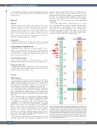
We identified 56 patients with homozygous (n=39) or compound heterozygous (n=17) mutations in MPL (Tables 1; Online Supplementary Table S1). We detected 38 different mutations (Figure 1, Table 2), 17 out of them are novel (Tables 1 and 2; Online Supplementary Table S2).
Six different nonsense mutations (allele frequency 20%; including three novel mutations) and three different frame shift deletions (allele frequency 13%) affected 20 different patients (Table 2A and B; Online Supplementary Table S2).
Five different splice site mutations (allele frequency 10%, two novel) affected 11 patients (ten families). With the exception of c.391+5G>C, all are predicted to lead to a complete loss of function (Table 2C). Prediction was confirmed for c.79+2T>A by measurement of missing CD110 surface expression on hematopoietic progenitors (Figure 2, see below) and for 213-1G>A and c.79+2T>A by the severe course of the disease in the affected patients. In contrast, patients with the mutation c.391+5G>C, allowing a residual natural splicing,26 had a less severe course and measurable CD110 expression on hematopoietic progenitors (Figure 2).
The majority of mutations in our patient cohort were missense mutations (24 different mutations in 35
patients, allele frequency 57%, 12 novel, Table 2D) Two hotspots (amino acids 102-104: 18 alleles, 12 patients; proline residues 135-136: six alleles, five patients) account for 21% of all mutated alleles. Fifteen of 24 missense mutations are predicted to be deleterious by all applied algorithms, 23 of 24 by at least one of the algorithms (Table 2D).
Due to the small number of individual cases it is diffi- cult to predict clinical courses from the individual mis- sense mutations. Specifically severe courses were observed in patients affected from p.Arg102Pro, p.Trp154Arg, and p.Leu169His. The latter one was found in three unrelated patients from Chile suggesting a founder mutation with regional significance.
Figure 1. MPL alleles in CAMT patients All MPL mutations found in our cohort of congenital amegakaryocytic thrombocytopenia (CAMT) patients are depicted on the left side beneath the exon structure of the MPL transcript with corresponding numbering of bases (coding sequence). Every symbol represents a mutated allele in one patient. The derived protein structure with the functional domains of the receptor protein and corresponding numbering of amino acids is shown on the right. Circles: missense mutations; diamonds: nonsense mutations; trian- gles: frame shift deletions; equal signs: splice site mutations. Mutations with less severe phenotypes are marked in green color (see main text). SP: signal pep- tide, TM: transmembrane domain
2440
haematologica | 2021; 106(9)
