
Page 68 - 2021_06-Haematologica-web
P. 68
E.R. Finch et al.
result in decreased osteonecrosis and would not interfere with the efficacy of antileukemic therapy.
Methods
Treatment
Experiments were approved by the Institutional Animal Care and Use Committee of St. Jude Children’s Research Hospital (SJCRH; Memphis, TN, USA): protocol numbers 423-100428 and 465-100549.
All mice received prophylactic antibiotics in drinking water (herein referred to as “base-water”): continuous tetracycline (1 g/L; Sigma-Aldrich, St. Louis, MO, USA) and intermittent sul- famethoxazole/trimethoprim oral suspension (600/120 mg/L for 3.5 days/week; Aurobindo Pharma, USA, Inc., Dayton, NJ, USA). Mice were randomized to receive continuous dexametha- sone (0.4 mg/mL sodium phosphate solution; Fresenius Kabi, Lake Zurich, IL, USA) in drinking water, herein referred to as “dexamethasone water” (3 mg/L in the osteonecrosis model and 4 mg/L in the BCR-ABL+ ALL model).21-23
The folic acid-deficient diet (0.2 ppm folic acid; TestDiet, Richmond, IN, USA) was the “base-diet”.21 Fenofibrate (Sigma Aldrich, St. Louis, MO, USA) was added to the base-diet for a final concentration of 0.2% fenofibrate (w/w) to make the “fenofibrate-supplemented diet” (TestDiet, Richmond, IN, USA). This dose had been used in prior experimental rodent models.24-26
Serum triglycerides were measured with an ABX Penta 400 instrument (Horiba, Montpellier, France) after the animals had fasted for 12 to 16 h (food was withheld; mice had free access to water).
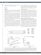
A
Mice were randomized to one of four treatment groups: con- trol (base-water and base-diet); fenofibrate-only (base-water and fenofibrate-supplemented diet); dexamethasone-only (dexam- ethasone-water and base-diet); or dexamethasone + fenofibrate (dexamethasone-water and fenofibrate-supplemented diet).
Osteonecrosis model
The protocol was modified from that previously described.21,22,27- 29 At postnatal days 26 to 28, male Balb/cJ mice (bred in-house at SJCRH) were randomized by body weight (13.5 g, 95% confi- dence interval [95% CI]: 13.0- 14.7) to 6 weeks of treatment. Fasting serum triglycerides were measured after 1, 3, and 6 weeks of treatment. At the end of treatment, white adipose tissue; and plasma dexamethasone and fenofibric acid levels were measured. Osteonecrosis and epiphyseal arteriopathy were evaluated and scored in both distal femora, by a board-certified veterinary pathologist (LJJ), as described previously21,22,27-29 Details are provid- ed in the Online Supplementary Methods.
BCR-ABL+ acute lymphoblastic leukemia model
The BCR-ABL (p185+, Arf-/-) luciferase-positive cell line (BCR- ABL+) was generated.23,30-32 Female 8-week-old matched syngene- ic mice (C57Bl/6J, Arf-wildtype; Jackson Laboratory, Bar Harbor, ME, USA) received intravenous injections of 2,000 BCR-ABL+ cells. Bioluminescent imaging was performed weekly,23 to mon- itor leukemic burden. At day 3, mice were stratified by lumines- cent signal and body weight to treatment (n=10/treatment group). Fasting serum triglycerides were measured at day 24. Treatment ended at day 28: mice were maintained on base- diet/base-water until a humane endpoint or the end of study at day 63. Details are given in the Online Supplementary Methods.
BC
Figure 1. Experimental design, body weight, and survival in dexamethasone-induced osteonecrosis. (A) Experimental design: male Balb/cJ mice were placed on 6 weeks of continuous treatment with dexamethasone, fenofibrate, or both. (B) Body weight relative to week 0 is shown as a median with 95% confidence interval (95% CI). Control mice and mice treated with fenofibrate (Feno) only weighed more than those treated with dexamethasone (Dex) only or Dex + Feno from weeks 1-6 of treatment (P<1x10⁻⁶). (C) Survival did not differ by treatment group. The survival rate was 100% in the control group (12/12) and the group treated with Dex + Feno (33/33), whereas it was 98% in both the Dex Only (23/24) and Feno Only (20/21) groups (P=0.55).
2096
haematologica | 2021; 106(8)
