
Page 40 - 2021_06-Haematologica-web
P. 40
M. Novakova et al.
Data analysis of genetic and immunophenotypic data
RNA-seq data (patients with switch, n=73; patients without switch, n=124), whole-exome sequencing (WES) data (patients with switch, n=30; patients without switch, n=70), single- nucleotide polymorphism (SNP) data (patients with switch, n=59; patients without switch, n=108) and ERG deletion data were ana- lyzed in diagnostic samples as published previously.24–26 Diagnostic samples were sorted if blasts comprised fewer than 80% of mononuclear cells. Purity of sorted populations was at least 90%. Patients in the B-other group without the following aberrations were assigned to the B-other rest subgroup: DUX4, ZNF384, MEF2D and NUTM1 rearrangements; BCR-ABL1-like and ETV6- RUNX1-like expression profiles; and iAMP21, PAX5-P80R and IKZF1-N159Y mutations.
In order to analyze RNA-seq and immunophenotypic data, uni- form manifold approximation and projection (UMAP)27 was used as the dimensionality reduction algorithm. Hierarchical clustering analysis (HCA) was performed using Euclidean distance and Ward's linkage.
Results
Incidence and features of patients with monocytic switch
Prospectively, we identified 61 patients with monocytic switch using the criteria described above (Table 1), which corresponded to 8% of patients.
No sex difference occurred in the monocytic switch (69% females vs. 56% males, not significant [n.s.]) but the mono- cytic switch was associated with older age at diagnosis
(median 7.8 vs. 4.5 years, respectively, P<0.001) and a high- er initial white blood cell (WBC) count (median 10,750/mL vs. 6,670/mL, P=0.038), lower hemoglobin level (median 7.8 g/dL vs. 8.8 g/dL, P=0.0046), higher platelet count (median 74,000/mL vs. 58,000/mL, P=0.048) and a higher proportion of blasts in PB (59% vs. 32%, P=0.0039), while the propor- tion in BM did not differ (91.2% vs. 90%, n.s.).
We confirmed the presence of patient-specific IG/TR rearrangements in the sorted monocytoid cells in 33 of 37 patients in whom the sorting was successful at various time points between d0 and d+33 (16 of 19 positive at d0; 23 of 27 positive between d+1 and d+14; 14 of 15 positive at d+15; eight of nine positive between d+16 and d+33). In the morphological examination of some patients, an increase in monocytic cells with variable morphology (monoblasts, promonocytes, and mature monocytes) was very clear. In three patients, at d+8 (DUX4r, n=2; and ZNF384r, n=1), we observed over 10,000 monocytes/mL in the PB samples (Online Supplementary Figure S3A and B).
Monocytic switch is most frequent in the PAX5-P80R, DUX4r, and ZNF384r genetic subtypes
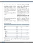
In order to study the relationship between monocytic switch and genetic background, routine (cyto)genetic inves- tigations were supplemented with a retrospective analysis using RNA-seq data, enabling a more detailed genomic characterization of the ALL patients. Patients with mono- cytic switch were unequally distributed across the ALL sub- types in the unselected consecutive cohort (chi square test P<0.0001; Table 1); they were significantly enriched in the PAX5-P80R-, DUX4r- and ZNF384r- -positive ALL subtypes
Table 1. Distribution of cases with monocytic switch in an unselected cohort of B-cell precursor acute lymphoblastic leukemia patients (n=726) stratified into genetic/biological subtypes.
Hypodi
BCP-ALL subtypes
Monocytic switch
P
P<0.0001
1 (1)
4 (12) ns
0 (0) ns 4 (24) ns 1 (11) ns 3 (6) ns
HHD1
ETV6-RUNX1 KMT2Ar TCF3-PBX1 BCR-ABL1
No n (%)
266 (97)
180 (99)
29 (88)
27 (100)
13 (76)
8 (89)
49 (94)
11 (27)
18 (95)
6 (60)
10 (100)
6 (100)
0 (0)
4 (100)
3 (100)
1 (100)
34 (100)
665 (92)
Yes n (%)
8 (3)
P<0.0001
ploidy2
B-other rest4 DUX4r BCR-ABL1-like ZNF384r ETV6-RUNX1-like iAMP21 PAX5-P80R NUTM1r
MEF2Dr IKZF1-N159Y Unknown5
30 (73)
1 (5) ns
P<0.0001
4 (40)
0 (0) ns 0 (0) ns
P=0.0022
5 (100)
0(0) ns 0(0) ns 0(0) ns 0(0) ns
61 (8)
P<0.0001
Total
1High hyperdiploidy with >50 chromosomes; 2<44 chromosomes; 3BCP-ALL negative for high hyperdiploid cases (HHD), ETV6-RUNX1, KMT2Ar, TCF3-PBX1, BCR-ABL1 and hypodiploidy; 4B-other analyzed by RNA sequencing (RNA-seq) and not belonging to any of the established subtypes; 5B-other not analyzed by RNA-seq (this subset is biased towards nonswitching cases because RNA-seq was performed in samples from all patients with monocytic switch,without identified genetic aberrations using polymerase chain reaction [PCR] and/or cytogenetics); 6P-value of the Fisher’s exact test on a comparison of the frequency of cases with monocytic switch in individual subsets vs. the frequency of switch among all the remaining cases. Multiple testing correction was done using Benjamini-Hochberg procedure. ns: no statistically significant difference; BCP-ALL: B-cell precursor acute lymphoblastic leukemia.
2068
haematologica | 2021; 106(8)
B-other3
