
Page 246 - 2021_06-Haematologica-web
P. 246
Letters to the Editor
One third of alloantibodies in patients with sickle cell disease transfused with African blood are missed by the standard red blood cell test panel
Studies on red blood cell (RBC) antibodies in Africa routinely use standard test cells from donors of Caucasian descent. There are no systematic data on alloimmunization against antigens that are almost exclu- sively present in Africans. We studied the prevalence of antibodies in transfused Ghanaian patients with sickle cell disease (SCD) using standard test cells (representing predominantly antigens more common in Caucasians (Caucasian antigens) and cells expressing antigens more common among Africans (African antigens). Antibodies were present in 16% of 221 patients; 31% of these were directed against African antigens that were not detected with standard test cells. Our findings are not only rele- vant for an African setting, but also for Western blood banks that are developing strategies to recruit more African donors.
Transfusions in patients with SCD are associated with high rates of red blood cell (RBC) alloimmunization against multiple antigens. Alloantibody screening is per- formed with standard reagent test cells, mostly from donors of Caucasian descent that lack antigens that are more prevalent in Africans. Alloimmunisation involving antigens that are almost exclusively present in Africans has not been studied systematically in a setting where both patients and donors are Africans. We determined the prevalence of RBC antibodies against Caucasian and African antigens in multi-transfused patients with SCD in Ghana, where pre-transfusion antibody screening and indirect antiglobulin crossmatch are not routine.
Our cross-sectional study recruited patients between July and December 2018, from two tertiary hospitals. Patients were eligible for inclusion if they were at least 2 years of age and had received at least two transfusions at least 6 weeks before study enrolment (to allow time to develop antibodies). Patients were episodically trans- fused with non-leucoreduced whole blood from African donors. Donors were not screened for sickle cells. Participants’ demographics and transfusion history were retrieved from hospital files. Patients or caretakers pro- vided this information, if missing from hospital files, using a standard questionnaire.
Plasma and buffy coat samples, taken at enrolment, were frozen at –80°C and transported to Sanquin, Amsterdam, the Netherlands, for routine antibody test- ing against a standard three-cell reagent panel (Bio-Rad Laboratories AG, Cressier, Switzerland), not expressing antigens that are more common in Africans, using a low ionic strength solution (LISS) indirect anti-globulin gel column agglutination test. Using the same method, anti- body identification was performed with commercial pan- els of reagent RBC of selected phenotypes and against eight selected cells with antigens that are very rare (<0.01% to 1%) in Caucasians but more frequent (0.5% to 32%) in Africans (i.e., MNS6 [He], MNS25 [Dantu], RH10 [V], RH20 [VS], RH30 [Goa], RH32, RH43 [Crawford] and KEL6 [Jsa]).1 These antigens were selected based on the availability of RBC expressing rare antigens archived in the Immunohaematology Diagnostics labora- tory at Sanquin. Antibody specificities were confirmed by re-testing with two RBC expressing and two RBC not expressing the target antigens. For patients with anti-D, RHD genotyping was done on genomic DNA by Multiplex Ligation-dependent Probe Amplification (MLPA) assay according to the manufacturers’ protocol
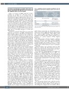
Table 1. Specificities of the 36 red blood cell antibodies in the 35 alloimmunized multi-transfused Ghanaian patients with sickle cell disease.
Blood group system
Rh
Kell
MNS Unidentified
Antibody specificity, (n)
Panel expressing antigens more common in Caucasians
E (10); D (7); G (2)
K (1)
s (1)
Pan-reactive (3);
Non-specified* (1)
African panel expressing antigens more common
in Africans
V/VS (2); VS (1); Goa (1); RH32 (1)
Dantu (3); He (2)
Non-specified* (1)
*The non-specified antibodies were probably against low frequency antigens. One patient had anti C+D.
(MRC Holland, Amsterdam, the Netherlands) using a thermocycler (Veriti, Applied Biosystems, Nieuwerkerk aan de IJssel, the Netherlands).2 When MLPA results were equivocal, DNA sequencing was performed to determine the RHD genotype. Sequence products were analyzed on a genetic analyzer (3730xl, Applied Biosystems).
The study was approved by the Committee on Human Research, Publication and Ethics, Kwame Nkrumah University of Science and Technology, and Korle Bu Teaching Hospital and Liverpool School of Tropical Medicine Institutional Review Boards. Patients or their caretakers gave written informed consent to participate in the study.
Statistical analyses were performed using the SPSS (IBM Corp., Armonk, NY, USA). Results for continuous variables were presented as medians (range) and categor- ical variables as frequencies (percentages).
We recruited 221 Ghanaian patients (123 women and 98 men; 89% hemoglobin [Hb] SS, 10% HbSC, one HbSD and one HbSb0-thalassemia). The median age at enrolment was 17 years (range, 2-66 years). Patients had received a median of three (range, 2-40) whole blood transfusions and the median period between last transfu- sion and study enrolment was 2.1 years (range, 6 weeks to 55.5 years).
Antibody screening, using standard test cells, was pos- itive in 24 patients (10.9%) and revealed 25 antibody specificities (Table 1). Although D antigen matching was routine in Ghana, anti-D was present in seven patients. RHD genotyping of these patients revealed that three women and two men had only RHD-null alleles (three RHD*01N.01/RHD*01N.01, one RHD*01 N.01/RHD*01N.03 and one RHD*08N.01/RHD*08N.01). In the three D negative (D-) women, anti-D could have been induced by a D positive (D+) pregnancy. For the two D- men, errors in blood group typing or mistakenly transfusing patients with D+ blood are possible causes for the anti-D. The presence of RH variants expressing weak D among D- African donors might have con- tributed to these mismatched transfusions. Two patients possessed variant RHD genes (RHD*03.04 and RHD*04.01) associated with D+ serology and carriers of these variants can make anti-D after D-antigen exposure.3,4
The nine patients with D, G and s antibodies and the three patients with pan-reactive antibodies, were not tested against the African antigens because African test cells lacking these antigens were not available. Of the
2274
haematologica | 2021; 106(8)
