
Page 201 - 2021_06-Haematologica-web
P. 201
Ars2 in the pathogenesis of DLBCL
BCR-expressing cell lines, but had no observable effect on DLBCL lines lacking Ars2-reactive BCR (Figure 4B). No toxic effect was observed with the control toxin LRPAP1-ETA’ against OCI-Ly3. Trypan blue staining after addition of 5 mg/mL Ars2-ETA’ showed that 35%, 2% and 0% of OCI-Ly3 cells were alive after 24 h, 48 h and 72 h, respectively, contrasting with findings for the wild-type HBL1 cell line without BCR reactivity against Ars2 (97% viable cells at 24 h; 96% at 48 h; and 97% at 72 h) (Figure 4B). In accordance with this, an increase of apoptotic cells was detected after incubation with Ars2- ETA’ in U2932 cells expressing Ars2-reactive BCR (Figure 4C), as demonstrated in the annexin V/propidium iodide assay.
Discussion
Beside the two relatively rare target antigens, ubiquiti- nated FamH83 and sumoylated JmJD4, in the present study, hypophosphorylated Ars2 was identified as a more frequent antigen of BCR from DLBCL lines and recombi- nant BCR from primary DLBCL cryospecimens. Ars2 is also known as serrate RNA effector molecule (SRRT). Its gene is located on chromosome 7q21 and the protein is a zinc finger protein consisting of 875 amino acids with a molecular weight of around 100 kDa. Ars2 was described as being involved in miRNA silencing by interacting with the nuclear cap binding complex,22 and as being involved in the innate immune response against RNA viruses by
AC
B
D
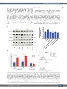
Figure 3. B-cell receptor pathway activation and induction of proliferation by Ars2. (A) B-cell receptor (BCR) pathway analysis by western blot in U2932 and HBL1 after addition of cognate/control antigens Ars2 or MAZ showed strong activation due to the addition of ARS2 to U2932 cells with upregulation of pTyr525/526 SYK, pTyr96 BLNK, pTyr759 PLCγ2 and pTyr223 BTK and higher expression of MYC. In contrast, no effect of ARS2 on the BCR pathway was observed in HBL1 cells. (B) Induction of proliferation by Ars2. Addition of Ars2 to OCI-Ly3 and U2932 lines resulted in a statistically significant (P<0.01: Student t-test) increase of proliferation, as determined by the EZ4U assay (columns represent formazan at an optical density [OD] of 450 nm), while addition of Ars2 had no effect on the TMD8 cell line. Columns and bars represent mean and standard deviation of three experiments. (C) Inhibition of Ars2-induced proliferation by neutralizing Ars2-reactive Fab. Addition of Ars2 together with Ars2-reactive (patient derived, case #4) recombinant Fab prevented induction of growth in U2932 cells. (D) Elevation of cytoplasmatic calcium levels by addition of Ars2. Flow cytometry analysis of cytoplasmic calcium levels using Fluo-4 dye showed an increase after addition of the cognate antigen ARS2 (blue) comparable to the effect of adding anti-IgM (black) to U2932 cells, but not after the addition of a control antigen MAZ (green). Addition of Ars2 to control the diffuse large B-cell lymphoma line HBL1 did not result in elevated calcium levels.
haematologica | 2021; 106(8)
2229
