
Page 200 - 2021_06-Haematologica-web
P. 200
L. Thurner et al.
nal FLAG tag in OCI-Ly10, OCI-Ly3 and, as a control, HBL1 (Figure 2D and Online Supplementary Figure S4).
Frequency, titers, and IgG subclasses of Ars2 serum antibodies
Ars2 antibodies were detected by ELISA in the sera of four of 98 patients with DLBCL, with titers ranging from 1:800 to 1:1600, and in one of 400 healthy controls. All four patients with Ars2 antibodies in their sera (#22, #27, #41, #73) were carriers of hypophosphorylated Ars2 in the cells of their peripheral blood (Figure 2B), but this iso- form was not detected in the peripheral blood of any of the 94 other patients, resulting in a statistically significant relationship between serum Ars2-autoantibodies and the presence of the hypophosphorylated Ars2 in peripheral blood (Fisher exact t-test: two-tailed P<0.0001).
Effects of Ars2 on diffuse large B-cell lymphoma lines
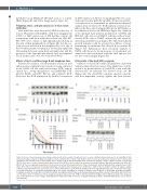
Western blot analysis of BCR pathway activation after addition of recombinant Ars2 revealed a strong activation in the U2932 cell line with Ars2-reactive BCR, demon- strated by a strong upregulation of pTyr525/526 SYK, pTyr96 BLNK, pTyr759 PLCγ2, and pTyr223 BTK. Moreover, this BCR stimulation by Ars2 led to increases
in MYC expression. However, regarding MYC two cases with Ars2-reactive BCR (#4 and #10) did not have MYC- overexpression, as determined by immunohistochemical analysis (data not shown). No BCR pathway activation was induced by the control antigen MAZ in the U2932 line or by addition of Ars2 to the HBL1 line (Figure 3A). Addition of recombinant Ars2 induced proliferation of U2932 and OCI-Ly3 cells, but not of DLBCL cell lines without Ars2- reactive BCR, such as TMD8, analyzed by the tetrazoli- um/formazan EZ4U assay (Figure 3B). This Ars2-induced growth stimulus could be reverted by addition of Ars2- neutralizing recombinant Fab derived from patient #4 (Figure 3C). Furthermore, flow cytometry analysis of U2932 cells showed a strong increase of cytoplasmic cal- cium levels after incubation with the Ars2 epitope, but not the control antigen (Figure 3D).
Cytotoxicity of the Ars2/ETA’ conjugate
Addition of Ars2-ETA’ resulted in inhibition of growth analyzed in proliferation assays. This inhibition could be reverted by preincubation of Ars2/ETA’ toxin with the Ars2-reactive recombinant Fab derived from case #4 (Figure 4A). The Ars2-ETA’ conjugate exerted a specific and dose-dependent toxicity against the Ars2-reactive
AC
BD
Figure 2. Ars2 is exclusively hypophosphorylated in patients with Ars2-reactivity of lymphoma B-cell receptors. (A) Western blot and isoelectric focusing (IEF) of Ars2 derived from diffuse large B-cell lymphoma (DLBCL) cell lines. Western blot of Ars2 in nine DLBCL cell lines revealed no difference in Ars2 between cell lines with and without Ars2-reactive B-cell receptors (BCR) (above). However, IEF of Ars2 in the DLBCL lines showed a less negative charge of Ars2 in OCI-Ly3, OCI-Ly10 and U2932. These three cell lines had exclusively Ars2-reactive BCR. (B) IEF of Ars2 of whole blood derived from DLBCL patients with Ars2-autoantibodies (#22, #27, #41, #73). The less negatively charged Ars2 isoform was also detected In the peripheral blood of these four patients. Ars2 autoantibody titers ranged between 1:800 and 1:1600 in these four patients. (C) Alkaline phosphatase treatment led to disappearance of differences in IEF of Ars2. A stronger reduction of negative charges of Ars2 by dephosphorylation was observed in cases/cell lines without Ars2-reactive BCR. (D) Identification of the hypophosphorylated sites by site-directed mutage- nesis and transfection of C-terminally FLAG-tagged Ars2 into U2932 and HBL1. In contrast to wild-type Ars2, mutations in Ser372Ala, Ser374Ala, Ser376Ala, Ser348Ala, Ser349 Ala, Ser357Ala, Ser361Ala, Ser365Ala, Ser368Ala and Ser370Ala resulted in hypophosphorylated Ars2 isoforms in both HBL1 and U2932; how- ever the Ars2 isoform of U2932 was still less negatively charged compared to that of HBL1. Only the mutants Ser328Ala and Ser341Ala resulted in the disappear- ance of this difference in electric charge, identifying both Ser328 and Ser341 as the sites of hypophosphorylation. Murine anti-FLAG-antibody was used as the pri- mary antibody.
2228
haematologica | 2021; 106(8)
