
Page 181 - 2021_06-Haematologica-web
P. 181
BMPR2 regulates the self-renewal of adult HSCs
KL
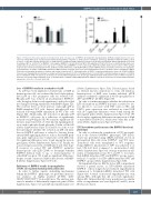
Figure 3. Reduced self-renewal capacity of hematopoietic stem cells upon loss of BMPR-II. (A) Competitive transplantation. (B) Representative fluorescence acti- vated cell sorting (FACS) plots of peripheral blood (PB) showing CD45.1 competitor vs. CD45.2 donor contribution, and lineage distribution within the CD45.2 subset of cells. T: T cells; B: B cells; M: myeloid cells. (C) CD45.2 donor contribution in PB of primary recipients at 16 weeks post transplant. Mean engraftment 50.6%±6.2 for wild-type (WT) vs. 36.5%±7.6 for BMPR-II-/-. Heterozygotes (BMPR-II+/-) did not differ from WT in PB engraftment (50.4%±7.1). Due to statistically detectable vari- ability between experiments, paired t-test was used to compare WT and BMPR-II-/- (n=4-7). (D) Lineage distribution within CD45.2 donor subset of PB in primary recipients at 16 weeks post transplant (n=4-7). (E) Representative FACS plots of LSK-SLAM CD45.1/2 stain of BM of primary recipients at 16 weeks post transplant. (F) Quantification of CD45.2+ LSK-SLAM populations in BM of primary recipients at 16 weeks post transplantation (n=4-7). (G) CD45.2 donor contribution in BM at 16 weeks post transplant in primary, secondary, and tertiary recipients (n=4-7). (H) Quantification of CD45.2+ Long-term hematopoietic stem cells (LT-HSC) in BM at 16 weeks post transplantation in primary, secondary, and tertiary recipients (n=4-7). (I) Transplantation of sorted LT-HSC to lethally irradiated recipients. (J) CD45.2 donor contribution in PB of recipients at 16 weeks post sorted LT-HSC transplant (n=6). (K) Lineage distribution within CD45.2 donor subset of PB in recipients at 16 weeks post sorted LT-HSC transplant (n=6). (L) Quantification of CD45.2+ LSK-SLAM populations in BM of recipients at 16 weeks post sorted LT-HSC transplan- tation (n=6). *P<0.05; **P<0.01; †P=0.078.
Loss of BMPR-II results in a reduction of p38
As p38 has been implicated downstream of BMP in hematopoietic cells, we evaluated the level of phosphory- lated p38 in c-kit+ progenitor cells by western blot. Phospho-p38 was reduced in un-stimulated BMPR-II-/- cells, though it did not reach significance, and its level did not change following stimulation with BMP4 (Figure 5D). We could not detect a robust increase of phospho-p38 in BMP4-stimulated WT cells. Instead, phospho-p38 was reduced following BMP4 stimulation in WT c-kit+ cells (Figure 5D). Additionally, the reduction of phospho-p38 in BMPR-II-/- cells may be a reflection of significantly reduced total p38 (Figure 5E). We found no significant dif- ferences in protein levels of other known signaling medi- ators such as phospho-Limk, phospho-Cofilin, or RhoA/B (Online Supplementary Figure S7A and B). In order to fur- ther investigate whether the reduction in LSK cell num- bers in BMPR-II null mice is related to known down- stream BMP signaling mediators such as the MAPK path- way, gene expression was evaluated in sorted LSK cells. No significant differences were found among the investi- gated genes (Online Supplementary Figure S7C). Finally, we assessed expression levels of BMP type-I and other type- II receptors in sorted WT and BMPR-II-/- primitive hematopoietic cells (LSK CD48-) to determine whether BMPR-II deletion leads to up- or down-regulation of other BMP receptors. We found no significant differences, despite a trend of increased Alk3 in the absence of BMPR-
II (Online Supplementary Figure S7D to E).
Deficiency of BMPR-II results in up-regulation of TJP1 in long-term hematopoietic stem cells
In order to further explore underlying mechanisms behind the BMPR-II-/- phenotype, we performed microar- ray analysis on highly purified LT-HSC (LSK- CD150+CD48-CD9hi)35 from adult mice. The analysis gen- erated a number of differentially expressed genes (Online Supplementary Figures S8 and S4B) and enriched gene sets
(Online Supplementary Figure S4A). Selected genes, based on relevant known connections to stem cell function, hematopoiesis or BMP, were further validated. qPCR analyses confirmed a significant 2.4-fold up-regulation of TJP1 in BMPR-II-/- LT-HSC (Figure 5F).
In order to further investigate whether the reduction in LSK cell numbers in BMPR-II null mice is related to fac- tors known to associate with TJP1 such as SRC and STAT3, gene expression was evaluated in sorted LSK cells. No significant differences were found among the investigated genes (Online Supplementary Figure S7C). We also found no significant differences in expression of Alpk or microRNA 15a/23b/27a, which were other hits in the array (Online Supplementary Figure S7F and G).
TJP1 knockdown partly rescues the BMPR-II knockout phenotype
In order to evaluate the contribution of TJP1 up-regula- tion to the observed BMPR-II-/- phenotype, TJP1 knock- down was performed using shRNA lentiviral vectors in ckit-enriched BM cells from BMPR-II-/- and WT mice. Transduced cells were transplanted to WT recipients. Using shRNA-C knockdown of TJP1 was achieved to at least 0.51-fold level (compared to un-transduced cells) (Online Supplementary Figure 9A). Average transduction effi- ciency at transplantation was 36 % and 33 % for scram- bled-shRNA transduced WT and BMPR-II-/- groups respec- tively; 21 % and 26 % for TJP1-shRNA transduced WT and BMPR-II-/- groups (Online Supplementary Figure S9B).
In transplanted mouse BM the donor LSK compartment showed a partial rescue, as TJP1-shRNA transduced BMPR-II-/- cells no longer showed reduced engraftment in comparison to Scrambled-shRNA transduced WT cells (Figure 6A). A trend of increased engraftment was seen among HSC, although this did not reach significance (Figure 6B). In hierarchically lower populations no similar effect on engraftment was seen (Figure 6C and D), nor in PB (data not shown).
haematologica | 2021; 106(8)
2209
