
Page 168 - 2021_06-Haematologica-web
P. 168
Z. Shah et al.
AB
C
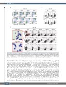
Figure 3. Blood cell populations are differently affected by the MYB deficiency depending on the stage of differentiation. (A) H1-isogenic MYB knockout cell lines develop normally on day 6 of differentiation. Total live differentiated cells were taken for the analysis, as in (B) and (C). Representative data of more than ten exper- iments are shown. (B) On day 10 the DKO cell lines demonstrate subtle differences in the CD43/CD45 cell staining. Representative data of three experiments are shown. (C) Day 20 phenotype of the MYB knockout cell lines. Representative data of six experiments are shown. Two cytospin panels on the left represent May- Grünwald staining of sorted CD66b+CD86- neutrophils and CD66b-CD86+ cells of the monocyte-macrophage lineage. Scale bar, 10mm.
Erythroid, Primitive). The ability of the primitive progeni- tors to form large colonies of erythroid cells suggests that the assay medium is optimal for analyzing primitive pro- genitors generated in the cytokine-free differentiation.
First, we determined that the vast majority of hematopoietic progenitors were MYB-Venus+ (Figure 5C). Of note, the analysis of the sorted cell populations demon- strated extremely high efficiency of hematopoietic pro- genitor generation in our differentiation system: up to 17% of all day 6 CD34+MYB-Venus+ blood cells generated CFU-C colonies in the assay medium. Then, we compared the generation of the progenitors by WT and mutant hESC lines on day 6 and day 10 of differentiation. Progenitors of higher proliferative potential, BFU-EP and CFU-MixP, were practically absent in differentiated cul- tures of MYB-null hESC, and the myeloid progenitors were also affected (Figure 5D). Furthermore, we observed the effects of MYB haploinsufficiency on the generation of
these progenitors from SKO cells. Only day 6 erythroid progenitors of low proliferation potential, CFU-EP, could be detected at a comparable density in the MYB-null cul- tures. However, CFU-EP in day 6 non-adherent DKO cell fractions were completely lost (Figure 5D). Moreover, instead of forming typical compact cell colonies, nearly all of day 6 MYB-null CFU-EP generated aberrant loose colonies, and the morphology of cells from those colonies was abnormal (Figure 5E). Almost all CFU-myeloid colonies generated by the DKO cells were composed exclusively of macrophages and were notably smaller compared to the WT and SKO counterparts (Figure 5F). The survival of the CFU-M subset of CFU-myeloid pro- genitors confirms the previously published data on MYB- independence of hPSC-derived macrophages and macrophage progenitors.10 Taken together, these observa- tions demonstrate that primitive erythroid and primitive mixed progenitors are non-functional in differentiated cul-
2196
haematologica | 2021; 106(8)
