
Page 122 - 2021_06-Haematologica-web
P. 122
S. Cordes et al.
and the endothelial monolayer was disrupted (Online Supplementary Figure S2D and E). During intestinal aGvHD, clotting was present in the colonic microvascula- ture as well as convolution of the endothelial monolayer (Online Supplementary Figure S2E). In conclusion, TEM revealed an extensive endothelial damage in target organs during aGvHD.
Pericyte coverage, tight junctions and endothelial leakiness during acute graft-versus-host disease
As a method to quantify endothelial damage in exper- imental aGvHD, we analyzed pericyte-coverage of CD31+ vessels in the colon and liver by fluorescence microscopy. Representative pictures are shown in Figure 3A and G. We found reduced pericyte-coverage of ves- sels in hepatic sinusoids (Figure 3B) and in colonic mucosa (Figure 3H) during aGvHD, indicating a dam- aged endothelial monolayer. In order to address the question whether the endothelial barrier function is altered during aGvHD, we assessed the endothelial
expression of the tight junction protein ZO-1 and the intercellular junction protein VE-cadherin in vessels of aGvHD target organs a shown in the representative pic- tures in Figure 3C and I. Immunostaining revealed reduced endothelial ZO-1 abundance during aGvHD, which correlates with reduced numbers of intact tight junction (Figure 3D and J). Moreover, colonic microvas- cular and hepatic sinusoidal endothelium VE-cadherin abundance was reduced (Figure 3E and K).
In order to investigate, if the reduced expression of tight junction and intercellular connection proteins of the endothelium during aGvHD have functional conse- quences in vivo, we injected Evans blue solution intra- venously and analyzed the penetration into the organs. In allo-HSCT recipients with aGvHD endothelial leakage was significantly increased in liver (Figure 3F) and colon (Figure 3L) as compared to HSCT recipients without aGvHD.
Next, we were interested if the detected endothelial changes occurred exclusively in the target organs during
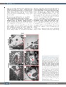
A
B
CD
EF
Figure 2. Visualization of acute graft-versus-host dis- ease-associated ultrastructural changes in the liver by transmission electron microscopy. Representative pictures of sections from liver taken at day+15 after experimental hematopoietic stem cell transplantation (HSCT) in the chemotherapy based B6→BDF model. (A and B) Liver sinusoidal endothelial monolayer after syngeneic-HSCT (syn-HSCT) without acute graft-ver- sus-host disease (aGvHD). (A) Normal, fenestrated sinusoidal blood vessel completely covered with endothelial monolayer. (B) Higher magnification of a 100 nm large fenestration of the endothelium in the liver. (C to-F) Sinusoidal liver endothelial monolayer after allo-HSCT during aGvHD. (C) Liver sinusoidal vessel with destroyed and unregularly shaped endothelial monolayer in contact with an immune cell. (D) Higher magnification of contact zone between immune cell and endothelial cell. (E) Blistering of the endothelial monolayer with a platelet in the region of injury. (F) Higher magnification of endothelial blistering. The perivascular space is marked by a red triangle. V: vessel lumen; EM: endothelial monolayer; F: fenestrated endothelium; IC: immune cell; E: erythrocyte; P: platelet; red circle: loss of endothelium; red triangle: endothelial blister- ing. Control groups (no aGvHD) were transplanted with the same bone marrow cell numbers and T-cell numbers from syngeneic donors.
2150
haematologica | 2021; 106(8)
