
Page 121 - 2021_06-Haematologica-web
P. 121
aGvHD impact on endothelium
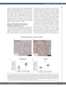
aGvHD. In contrast, Casp3+ EC were frequent in intestin- al biopsies of patients diagnosed for grade III-IV aGvHD. Figure 1 shows exemplary pictures of colon sections in human allo-HSCT recipients without aGvHD (Figure 1A) versus grade III-IV intestinal aGvHD (Figure 1B). Quantification revealed a significant increase in percent- age of Casp3+ vessels in duodenal (Figure 1C) and colonic mucosa (Figure 1D) of grade III-IV aGvHD. Patient char- acteristics and clinical data are given in the Online Supplementary Table S1 (for Figure 1C) and the Online Supplementary Table S2 (for Figure 1D). Our data demon- strate endothelial apoptosis during severe intestinal aGvHD in human allo-HSCT recipients.
Micro-structural endothelial changes during experimental acute graft-versus-host disease in transmission electron microscopy
In order to further investigate micro-structural changes of the endothelium we used experimental aGvHD mod- els. We first performed transmission electron microscopy (TEM) of liver (Figure 2A to F) and colon (Online Supplementary Figure S2A to F) at day+15 after allo-HSCT. We found that the hepatic sinusoidal endothelium in allo-
AB
HSCT recipients without aGvHD is not affected. The endothelial monolayer as well as the EC-cell contacts were intact (Figure 2A and B), and we observed a normal endothelial monolayer in hepatic sinusoids (Figure 2B). In contrast, the hepatic sinusoidal endothelium during aGvHD was severely damaged with close immune cell- EC interactions (Figure 2C). Furthermore, we observed a discontinuous endothelial monolayer at the EC-immune cell contact zone during hepatic aGvHD (Figure 2D). In addition, we found blistering of the endothelial monolay- er (Figure 2E) as well as platelet adhesion on the sinu- soidal endothelial monolayer (Figure 2E) and in the blis- tered endothelium (Figure 2F) during hepatic aGvHD. In colonic mucosa, vessels were again normal in allo-HSCT recipients without aGvHD (Online Supplementary Figure S2A to F). The endothelial monolayer was well-struc- tured, smooth and surrounded by pericytes (Online Supplementary Figure S2A) with intact tight junctions (Online Supplementary Figure S2B). In contrast, during intestinal aGvHD the endothelial monolayer was ruffled (Online Supplementary Figure S2C) with perivascular fib- rinogen deposits (Online Supplementary Figure S2A, C, D and F). Endothelial cytoplasm was enriched with vesicles
CD
Figure 1. Endothelial damage in human intestinal biopsies. (A) Exemplary picture of a colon biopsy of a patient after allogeneic hematopoietic stem cell transplan- tation (allo-HSCT) without histologic evidence of acute graft-versus-host disease (aGvHD) and low level of endothelial apoptosis. The white dotted lines indicate ves- sel lumen and the arrow indicates one apoptotic caspase 3 positive (Casp3+) endothelial cell. (B) Exemplary picture of a colon biopsy of a patient with grade III-IV intestinal aGvHD and increased endothelial apoptosis. The white dotted lines indicate vessel lumen and the arrows indicate apoptotic Casp3+ endothelial cells. (C) Quantification of Casp3+ events in duodenal endothelium of allo-HSCT recipients given in percent of vessels in high-power fields (HPF). (D) Quantification of Casp3+ events in colonic endothelium of allo-HSCT recipients given in percent of vessels in HPF. Percentage of Casp3+ vessels was tested for significance by Student’s t-test (***P<0.001; n=7-11 patients per group). Error bars indicate mean ± standard error of the mean.
haematologica | 2021; 106(8)
2149
