
Page 264 - 2021_07-Haematologica-web
P. 264
Case Reports
Light chain proteinuria revealing mu-heavy chain disease: an atypical presentation of Waldenström macroglobulinemia in two cases
Heavy chain diseases (HCD) are rare mature B-cells proliferative disorders first described in 19641 and charac- terized by the production of a paraprotein consisting of truncated heavy chains devoid of bound light chains. Normal immunoglobulin is composed of two heavy chains and two light chains joined by disulfide bonds at the heavy chain constant domain 1 (CH1). In the absence of light chains, the heat shock protein BiP binds to CH1 and retains the heavy chain in the endoplasmic reticu- lum.2 In HCD various mutations are responsible of splic- ing error leading to complete or partial deletion of CH1 and preventing therefore the binding of heavy chains to light chains as well as BiP.3,4 Three HCD involving the main immunoglobulin (Ig) classes have been described: a-HCD, γ-HCD and m-HCD which is the least common. A single case of d-HCD has been reported. m-HCD is often associated with a B-cell lymphoid disorder such as chronic lymphocytic leukemia with hepatosplenomegaly. It has also been described in association with myelodys- plasia, cirrhosis and auto-immune disease.5
Waldenström macroglobulinemia (WM) is a lympho- plasmacytic lymphoma secreting monoclonal IgM, main- ly κ, which is strongly associated with the MYD88 L265P somatic mutation.6 Patients may be asymptomatic or may present symptoms related either to bone marrow infiltration and/or to IgM gammopathy physico-chemical properties (including hyperviscosity, auto-immune
hemolytic anemia, cryoglobulinemia, anti-MAG neu- ropathy). Serum-free light chains (sFLC) rarely reach high levels in WM and complications related to light chains, like nephropathy or amyloidosis, are uncommon7 com- pared to multiple myeloma.
We report here two cases with IgM κ monoclonal gam- mopathy visible as a small peak on serum protein elec- trophoresis (SPEP) and in contrast to a high level of sFLC revealing m-HCD associated with WM.
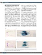
Case 1. In December 2018, a 79 year-old man with chronic renal failure of unknown cause presented with acute renal failure (creatinine 1,160 mmol/L) associated with nephrotic syndrome (proteinuria 2,9 g/24 hours and albumin 27 g/L) leading to end-stage renal failure requiring hemodialysis and normocytic non-regenerative anemia. The blood count was as following: hemoglobin 6 g/dL, platelets 208,000/mm3, neutrophils 3,100/mm3, lymphocytes 900/mm3. Kidney biopsy showed intersti- tial fibrosis and tubular atrophy associated with linear Congo red-negative deposits of κ-light chains along the basement membrane suggestive of Randall-type mono- clonal immunoglobulin deposition disease (MIDD). sFLC-κ were elevated at 2,585 mg/L with a κ/l ratio of 57/36. γ globulins were at 6,5 g/L and no peak was detected on SPEP but immunofixation was positive for monoclonal IgM κ (Figure 1A). Serum immuno-selection confirmed the presence of m-heavy chain (m-HC) (Figure 1B). Bone marrow aspiration and biopsy showed lym- pho-plasmocytic infiltration with 19% of lymphoid and plasma cells on aspiration and 25% of CD19+ CD20+ cells with monotypic expression of κ-light chain on flow cytometry. Immuno-histochemistry on biopsy identified
AB
CD
2034
Figure 1. Immunological tests. On the left side, serum protein electrophoresis and immuno-fixation for patient 1 (A) and patient 2 (C) who had both monoclonal IgM κ. On the right side, immuno-electrophoresis with immuno-selection for patient 1 (B) and patient 2 (D). Black arrow indicate precipitine line consisting of m-heavy chain (continuus arrow for patients, dotted arrow for positive control)
haematologica | 2021; 106(7)
