
Page 261 - 2021_07-Haematologica-web
P. 261
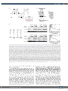
Figure 1. ALAS2 iron-responsive element mutation as modifier for erythropoietic protoporphyria clinical severity. (A) Pedigree of the studied family. Filled black symbol indicates the patient with clinical overt erythropoietic protoporphyria (EPP). Grey symbols indicate relatives (II.2 and II.4) with protoporphyrin IX (PPIX) over-production in red blood cells (RBC) associated with mild photosensitivity without EPP; the grandmother (I.1) presented with a suspected mild photosensi- tivity but no biochemical data were available; barred symbols indicate deceased subjects. Asterisks indicate subjects characterized at the molecular level. Partial chromatograms of ALAS2 iron-responsive element (IRE) sequences where the -38T>C mutation is located. Reference sequence for ALAS2: NM_000032.4. (B) Non-radioactive competitive electrophoretic mobility shift assays (EMSA) with wild-type (wt) and mutated (mut) ALAS2 IRE. Graphic representation of the ferritin H (FTH1) IRE sequences (nucleotides 60-85 in NM_002032.2) wt or mut -165ΔC, ALAS2 IRE sequence (nucleotides 93-125 in NM_000032.4) wt or the -38U>C mut, used for the synthesis of RNA probes in competitive EMSA experiment. RNA Watson-Crick pairs are depicted as A-U; U-A; C-G or G-C; wobble pairings are shown as U.G and not possible pairings are depicted as CxA. Fluorescent-labeled FTH1 IRE wt probe was incubated with increasing molar excess (2x, 5x, 10x, 20x, 40x and 80x) of unlabeled competitors corresponding to the FTH1 IRE wt type sequence (lanes 3-8; lanes 31-26), or the FTH1 IRE mutant -165ΔC (lanes 9-14; lanes 37-42), ALAS2 IRE wt sequence (lanes 17-22; lanes 37-42) or the ALAS2 -38U>C mutation (lanes 23-28; lanes 51-56). Samples were incubated with recombinant IRP1 (upper panel) or IRP2 (bottom panel) and resolved on acrylamide gels. Quantification of the signals of the shifted bands was performed using the Odyssey Infrared Imaging System (LI-COR Biosciences) and compared to the signal in lane N, set as 100%. Means ± standard deviation of at least three independent experiments. Statistical analysis by Student’s t-test (two-tailed) compares the signal given by the mutated IRE of FTH1 or ALAS2 sequences to the signal given by the corresponding wt IRE sequences, at each molar concentration *P<0.05, **P<0.01, ***P<0.001. F: free probe; N: no competitor added. (C) Proteins from CD34+ erythroid cells from proband’s mother (II.5) and the proband (III.2) cultured and differentiated over 14 days were extracted. Twenty micrograms were separated (4-12% NuPAGE gel) with MES NuPAGE buffer. Proteins were transferred to nitrocellulose membrane. ALAS2 was detected by chemiluminescence using specific antibody. The lower band is ALAS2 whereas the upper band, indicated with an asterix, is an unknown non-specific protein (Agios Pharmaceutical). The membrane was then probed with an anti-human beta-actin antibody as a loading control.
Case Reports
AC
B
nucleotide substitution (c.1102G>A; p.G298D) at the exon 7 of CLPX gene.11
The proband did not harbor mutations in the 11th exon of ALAS2 gene demonstrating that she is affected by an unusual form of EPP. Sequencing of the rest of the ALAS2 gene revealed an heterozygous T>C change at position -38 in the first exon, at the 5’ UTR and located inside the ALAS2 IRE motif (NM_000032.4; c.[- 38T>C];[=]) (Figure 1A and B). The mutation is absent in the 6,500 sequenced exomes in the Exome Variant Server database, excluding the possibility of being a neutral polymorphism. This variation was predicted to disturb the stability of the IRE, since the IRE position below the C8 bulge is critical to maintain closed the lower stem (IRE ALAS2 wild-tytpe [wt] predicted mini- mum free energy ΔG=-7.00 Kcal/mol vs. IRE ALAS2 mutant predicted minimum free energy ΔG=-5.60
Kcal/mol).12 In order to confirm the functional conse- quence of the -38U>C change in ALAS2 IRE, we per- formed non-radioactive competitive electrophoretic mobility shift assays (EMSA) with recombinant IRP1 or IRP213 (Figure 1B). As expected, the ferritin H (FTH1) wt positive control showed efficient competition, while its corresponding negative control, the FTH1 mutant - 165ΔC could not compete with the labeled FTH1 wt probe (Figure 1B, compare lanes 3-8 to 9-14 in upper panel and 31-36 to 37-42 in bottom panel). In addition, the ALAS2 -38U>C mutation totally abolished the abili- ty of the ALAS2 IRE to compete with FTH1 wt probe for IRP1 or IRP2 binding, in contrast to the wt ALAS2 IRE sequence (Figure 1B, compare lanes 17-22 to 23-28 in upper panel and 45-50 to 51-56 in bottom panel). Therefore, the -38U>C mutation in ALAS2 IRE abolish- es IRP1 and IRP2 binding affinity in vitro, which would
haematologica | 2021; 106(7)
2031
