
Page 250 - 2021_07-Haematologica-web
P. 250
Letters to the Editor
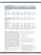
Table 2. Association between troponin elevation and other variables, and between coronary microvascular disease on cardiac magnetic res- onance imaging and other variables.
Clinical variables, number (%)
ACS at time of event No
Yes
AKI at the time event No
Yes
EKG changes No
Yes missing
OME use 24 hour prior to peak troponin elevation, median (range) missing
OME use 24 hour after peak troponin elevation,
median (range)
Normal Troponin (n=21)
19 (90) 2 (10)
Troponin data (N=63)† Elevated P*
Troponin (n=42)
25 (60) 0.02 17 (40)
CMD (n=15)
12 (80) 3 (20)
Cardiac MRI data (N=47)† Non-CMD P*
(n=32)
26 (81) 1.00 6 (19)
21(100) 24(57) 0.0002 NA NA
0 (0)
5 (100) 0 (0) 16
75 (75-75) 20
NA
18 (43)
27(73) 0.31 10(27)
5
64 (0-1524) 1.00 19
119 (0-3200) NA
7 (78) 2 (22) 6
NA
NA
11 (85) 1.00 2 (15)
19
NA
NA
Lab variables, median (range)
WBC count (×109/L) 12.2 (5.2-61) 13.7 (4.5-34.7) 0.39 11.9 (8-61) 13.6 (5.2-26.8) 0.35
Hemoglobin (g/dL) 7.7 (5.6-12) 7.7 (2.9-12.6) 0.93 7.8 (5.6-11.9) 7.6 (2.9-12) 0.34 Platelet count (×109/L) 272 (127-633) 165.5 (41-824) 0.007 282 (140-664) 259 (119-633) 0.30
Serum creatinine (mg/dL) 0.7 (0.1-1.4) 1.4 (0.4-7.6) 0.0002 0.8 (0.5-1.6) 0.7 (0.1-7.6) 0.41 LDH (U/L) 391.5 (210-1584) 498 (142-18500) 0.37 375 (142-1066) 486.5 (210-2647) 0.29
2020
CMD: coronary microvascular disease; MRI: magnetic resonance imaging; CMR: cardiac MRI; ACS: acute chest syndrome; AKI: acute kidney injury; EKG: electrocardiogra- phy; OME: oral morphine equivalents; WBC: white blood cell; LDH: lactate dehydrogenase; NA: not applicable; n: number. *Fisher’s exact test for categorical variables; Wilcoxon rank sum test for continuous variables.The number in parenthesis represents percentage of total patients for categorical variables and range for continuous vari- ables, which are reported as median. †Troponin level was not available for six of 69 patients and CMR was not available for 22 of 69 patients.
microvascular disease management in SCD patients. Patients are usually managed with standard acute coro- nary syndrome management with anticoagulation and antiplatelet agents. In patients with microvascular ischemia in general, aspirin, nitrate, beta-blockers, statins, and other coronary artery disease management interventions have been used but specific data for SCD is not available.13 Exchange transfusion with a goal HbS <30% has been reported to be effective in a patient with recurrent myocardial infarction.14 In a recent study evalu- ating the effect of hydroxyurea on skeletal and cardiac muscles, SCD patients treated with hydroxyurea had higher resting myocardial perfusion as compared to those without hydroxyurea.15 Newer therapies that target Hb modification could also have a role in the management, but prospective trials are needed to assess the effective- ness of various treatment strategies. We suggest that car- diac MRI with stress testing can be considered as a screening tool for patients with evidence of myocardial damage and recurrent chest pain. This could potentially identify patients who need to be started on medical ther- apy or considered for exchange transfusions. Improving access to cardiac MRI will be essential since a lack of its universal availability would be a hurdle in adopting this practice widely.
Kiranveer Kaur,1 Ying Huang,2 Subha V. Raman,3 Eric Kraut2 and Payal Desai2
1Division of Medical Oncology, The Ohio State University Wexner Medical Center, Columbus, OH; 2Division of
Hematology, The Ohio State University Wexner Medical Center, Columbus, OH and 3Krannert Institute of Cardiology, Indiana University School of Medicine, Indianapolis, IN, USA
Correspondence: PAYAL DESAI - payal.desai@osumc.edu doi:10.3324/haematol.2020.271254
Received: September 2, 2020.
Accepted: January 12, 2021.
Pre-published: January 21, 2021.
Disclosures: no conflicts of interest to disclose.
Contributions: PD is the principal investigator of this study and developed the study design, contributed to data interpretation, reviewed, and edited the manuscript; KK contributed to the data collection, analysis, interpretation, and manuscript writing;
YH contributed to the statistical analysis and interpretation of the data; SR and EH contributed to study design and manuscript editing.
References
1. Gladwin MT, Sachdev V. Cardiovascular abnormalities in sickle cell disease. J Am Coll Cardiol. 2012;59(13):1123-1133.
2. Martin CR, Johnson CS, Cobb C, et al. Myocardial infarction in sick- le cell disease. J Natl Med Assoc. 1996;88(7):428-432.
3. Raman SV, Simonetti OP, Cataland SR, et al. Myocardial ischemia and right ventricular dysfunction in adult patients with sickle cell disease. Haematologica. 2006;91(10):1329-1335.
4. Aslam AK, Rodriguez C, Aslam AF, et al. Cardiac troponin I in sickle cell crisis. Int J Cardiol. 2009;133(1):138-139.
5. Lippi G, De Franceschi L, Salvagno GL, et al. Cardiac troponin T dur- ing sickle cell crisis. Int J Cardiol. 2009;136(3):357-358.
haematologica | 2021; 106(7)
