
Page 248 - 2021_07-Haematologica-web
P. 248
Letters to the Editor
Myocardial injury and coronary microvascular
disease in sickle cell disease
Myocardial infarction and microvascular ischemic damage in the heart are one of the least well-described entities in the sickle cell disease (SCD) spectrum.1 Over the last few decades, some autopsy studies and case reports have described myocardial infarction without obstructive coronary artery disease suggesting microvas- cular ischemic injury in SCD patients.2 Impaired myocar- dial perfusion reserve in SCD has been demonstrated using different modalities including contrast echocardio- graphy, nuclear myocardial perfusion scans, single-pho- ton emission computerized tomography (SPECT) as well as cardiac magnetic resonance imaging (MRI).3 During a state of physiological stress or acute crisis, it can lead to myocardial injury which can be detected by serum tro- ponin measurement. The role of troponin in microvascu- lar disease in sickle cell disease has not been well defined. troponin-I was elevated (>0.4 ng/mL) in two of 32 patients 24 hours after the onset of acute crisis, with chest pain and electrocardiogram findings of sinus tachy- cardia and non-specific ST-T wave changes.4 In another study of six patients, troponin-T was normal 24 hours after admission for a sickle cell crisis in all patients.5
Cardiac magnetic resonance (CMR) is a non-invasive diagnostic tool that can be used for myocardial tissue characterization and assessment of coronary microvascu- lar disease (CMD).6 Due to high spatial resolution with first-pass perfusion imaging, it can visualize diffuse subendocardial perfusion abnormality resulting from CMD during the administration of a vasodilating drug such as adenosine.7 Late gadolinium enhancement (LGE) imaging visualizes decreased clearance of gadolinium- based contrast agents from the extracellular space in areas of damaged myocardium. LGE has become the in
vivo gold standard for visualization of myocardial injury from a variety of causes;8 subendocardial enhancement indicates an ischemic injury, whereas midwall enhance- ment indicates fibrosis of non-ischemic myocardial dis- ease and epicardial enhancement is consistent with inflammatory damage.9 Our study aims to estimate the prevalence of myocardial injury defined by elevated tro- ponin-I levels. We will also define the prevalence of coro- nary microvascular disease and other myocardial abnor- malities in an SCD cohort clinically referred for cardiac MRI.
We conducted a retrospective study of the SCD patients seen at The Ohio State University Wexner Medical Center over a period of 10 years from July 2005 to July 2015. Patients age 18 years or above, with tro- ponin-I level elevation (level >0.11 ng/mL) and/or cardiac MRI were included in the initial cohort. Coronary microvascular disease (CMD) on CMR was defined by the presence of either subendocardial damage by LGE or impaired myocardial perfusion by adenosine stress perfu- sion imaging. All other abnormalities were categorized as non-CMD due to a lack of specificity. Clinical and labo- ratory variables closest to the peak troponin elevation and cardiac MRI were recorded, if available within 4 weeks. For all patients with troponin level measurement and cardiac MRI, the date of death confirmed by chart review by June 2019 was recorded.
Out of 373 SCD patients, 69 had either troponin level measurement or cardiac MRI, or both done. The median age was 34 years (range, 19-67 years) with 30% of patients over the age of 40 years. Thirty-four (49%) patients were female. Seventy-five percent of the patients were hemoglobin (Hb) SS, and the rest 25% were other genotypes (SC 15%, S-βthal 9%). Median baseline Hb (defined by Hb level at a steady-state within the preced- ing year) was 8.0 g/dL (range, 4.5-13 g/dL) and median current Hb at the time of the event (either troponin ele-
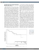
Figure 1. Overall survival comparison between coronary microvascular disease and non-CMD group. CI: Confidence Interval; Y: CMD present; N: no CMD; NE: not estimable.
2018
haematologica | 2021; 106(7)
