
Page 228 - 2021_07-Haematologica-web
P. 228
Letters to the Editor
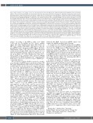
Figure 2. MLL contributes to the loading of let-7a onto AGO1 through interacting with RAN. (A) List of MLL-associated proteins identified by mass spectrometric analysis. 293T cells transfected with MLL were harvested and subjected to the nuclear-cytoplasmic fractionation. The cytoplasmic fractions were prepared for the immunoprecipitation assays followed by mass spectrometric analysis. (B) 293T cell lysates were treated with RNase A followed by anti-MLL immunoprecipitation. Western blots were performed using the indicated antibodies. (C) Direct interaction between MLLC180 and GST-RAN was examined. Left panels: western blots showing the inputs of purified GST-RAN and Myc-MLLC180. Right panels: the pull-down immunoblots were shown with GST-RAN as the bait and the pulled MLLC180 detected by an anti-Myc antibody. (D) 293T cells were untreated (upper panels) or treated with arsenite (0.5 mM, 45 min) (lower panels), then fixed and stained with the indi- cated antibodies. Note that eIF3 is specific for stress granules. Arrowheads show the localization of MLL with RAN and eIF3. Scale bar, 5 mm. (E) The RPISeq tool was used to predict the interactions between RAN and let-7a or pre-let-7a. The random forest (RF) classifier and support vector machine (SVM) classifier represent the confidence of the prediction. In performance evaluation experiments, predictions with probabilities >0.5 were considered “positive”. (F) 293T cellular lysates were prepared and anti-RAN RIP experiments were performed. Pulled down RNA were isolated, pre-let-7a and mature let-7a were analyzed by qRT-PCR using specific primers. (G) 293T cellular lysates were subjected to biotinylated- let-7a RNA pull-down assays. Then let-7a-immunoprecipitated RAN proteins were subjected to west- ern blot analysis. Scrambled miRNA were used as negative controls. (H) 293T-shScr and shRAN cells transfected with Agomir-negative control (NC) or Agomir- let- 7a mimic (let-7a) were subjected to dual luciferase reporter assays. The ratio of luciferase activity was measured and normalized to the value of the cells transfected with the control reporter and NC. (I) Extracts of 293T-shScr and shRAN cells, with the latter being rescued by shRNA-resistant RAN, were subjected to anti-AGO1 RIP assays. Pulled-down RNA were analyzed by qRT-PCR using specific primers for let-7a. (J) 293T-shScr and 293T-shRAN cellular lysates were subjected to biotinylat- ed-let-7a RNA pull-down assays. Then let-7a-immunoprecipitated AGO1 proteins were subjected to western blot analysis. Scrambled miRNA were used as negative controls. (K) 293T cell lysates were treated with RNase A followed by anti-AGO1 immunoprecipitation. Western blots were performed using the indicated antibodies. (L) Extracts of 293T-shScr and 293T-shMLL cells were collected and co-immunoprecipitation assays were performed and analyzed using the indicated antibodies. (M) The proposed mechanism through which MLL and RAN are involved in the loading of let-7a onto AGO1. MLL is required for the loading of let-7a onto AGO1 via a direct interaction with RAN. Thus, RAN serves as a molecular adaptor for the assembly of MLL-associated miRISC. NS, no significant difference. *P<0.05, **P<0.01, ***P<0.001. Data represent the mean and standared error of mean of three independent experiments.
plasm.12 According to the RPISeq online tool,13 RAN could bind the pre-miRNA and the mature miRNA (Figure 2E, Online Supplementary Figure S2A). Next, our RIP assay confirmed that RAN could bind not only the pre-miRNA but also the mature miRNA (Figure 2F, Online Supplementary Figure S2B). Moreover, RNA pull-down results showed a higher RAN expression in the let-7a or miR-10a-biotinylated group compared to that in the con- trol group (Figure 2G, Online Supplementary Figure S2C). These results were consistent with a previous finding that RAN was an RNA-binding protein,14 suggesting that RAN may be involved in the later steps of miRNA pro- cessing and function.
We next probed whether RAN is required to mediate gene silencing of miRNA targets. As shown in Figure 2H and Online Supplementary Figure S2D,E, luciferase activity in RAN-depleted cells was increased compared with that in control cells, indicating that the loss of RAN impaired the let-7a and miR-10a silencing functions. RIP experi- ments showed that the binding of both let-7a, miR-10a and MYC, HOXA1 to AGO1 was decreased in RAN- depleted cells, an effect that could be recovered by the reintroduction of RAN (Figure 2I, Online Supplementary Figure S2F-I). Our previous studies demonstrated that MLLC180 plays a causal role in the miRNA functional defi- ciency,3,4 so we then investigated the role of RAN-binding in MLLC180-regulated miRNA function. We found that MLLC180 failed to rescue the miRNA activity when Ran was depleted, an effect that could be recovered by MLLC180 together with reintroduction of RAN, suggesting that RAN was required for the MLLC180-mediated miRNA regulation (Online Supplementary Figure S2J-K). Given the fact that RAN is a small GTPase involved in nucleocyto- plasmic transport,10 we determined whether the GTPase activity of RAN is required for the functional interaction of the MLL-miRISC complex. As revealed in Online Supplementary Figure S2L, both wild-type RAN (RANWT) and GTPase-deficient mutant (RANQ69L) could partially reverse the deficits in the binding of let-7a to AGO1 caused by loss of endogenous RAN. We also observed that depletion of RAN significantly impaired the interac- tion between MLL and AGO1, which could be recovered by RANWT or RANQ69L re-expression (Online Supplementary Figure S2M), suggesting that the GTPase activity of RAN was not required for the function of the MLL-miRISC complex. Additionally, the binding of AGO1 to let-7a or miR-10a was decreased in RAN-depleted cells as revealed by a pull-down assay using biotinylated let-7a or miR-10a (Figure 2J, Online Supplementary Figure S2N). These results
indicated that RAN, beyond pre-miRNA export, was required for miRNA-mediated gene silencing.
To decipher the role of RAN in the function of miRISC, we tested the interaction between RAN and AGO1. We observed that AGO1 had an RNA-dependent indirect interaction with RAN (Figure 2K). Importantly, co- immunoprecipitation experiments revealed that besides AGO1, DDX6 a key P-body protein specifically involved in miRNA-mediated translational repression,15 interacts with RAN, but these interactions decreased significantly upon MLL depletion (Figure 2L), indicating that MLL is accountable for these interactions.
To further strengthen our findings, we explored how RAN behaves in a leukemic context. Co-immunoprecipi- tation assays performed in three leukemia cell lines, JM1, REH and U937, showed that MLL interacts with RAN (Online Supplementary Figure S2O). In REH and U937 cells, MLL together with RAN co-localized to stress gran- ules following arsenite treatment, as illustrated by immunofluorescence assay (Online Supplementary Figure S2P). Additionally, we found that the binding of let-7a to AGO1 was decreased in RAN-depleted REH and U937 cells, an effect that could be restored by the reintroduc- tion of RAN (Online Supplementary Figure S2Q-R). Consistent with the results obtained from 293T cells, we observed that AGO1 had an RNA-dependent indirect interaction with RAN in REH cells (Online Supplementary Figure S2S). Moreover, the interaction between AGO1 and RAN was impaired in MLL-depleted REH cells (Online Supplementary Figure S2T-V). As expected, the binding of RAN to AGO1 was reduced in MLL leukemic cells due to the downregulation of MLLC180 (Online Supplementary Figure S2W).
Collectively, in the present study, we demonstrated that MLL was required for recruiting let-7a and its target mRNA to the miRISC, partly through its direct binding partner RAN (Figure 2M), unraveling an unexpected role for RAN in the loading of miRNA onto AGO1. Our find- ings provide an alternate mechanism and expanded the functional scope of RAN in the miRNA processing path- way. Thus, the discovery of interplay between MLL and miRNA represents a new regulatory layer, and an addi- tional level of complexity, in the control of gene expres- sion.
Shouhai Zhu,1,2* Zhihong Chen,1* Dan Jiang,3*
Ruiheng Wang,1 Xiaoyan Cheng,1 Dan Li,1 Qiongyu Xv,1 Fei Zhao,2 Wootae Kim,2 Guijie Guo,2 Chunjun Zhao,1 Zhenkun Lou2 and Han Liu1
1998
haematologica | 2021; 106(7)
