
Page 226 - 2021_07-Haematologica-web
P. 226
Letters to the Editor
AC
B
DEF
GHI
JK
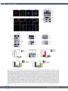
Figure 1. MLL is required for the loading of let-7a onto AGO1. (A) 293T cells were transfected with GFP-AGO1. Immunofluorescence experiments were per- formed to visualize the localization of GFP-AGO1 and MLL. MLL-CT antibody, which recognizes MLLC180 (aa2829-2883), was used to detect MLL. Scale bar, 5 mm. (B) Mll wild-type (Mll+/+) and Mll knockout (Mll-/-) MEF cells were transfected with GFP-AGO1. Immunofluorescence experiments were performed to visualize the localization of GFP-AGO1 and MLL. Arrowheads show the localization of MLL with the GFP-AGO1. Scale bar, 5 mm. (C) 293T cells were transfected with FLAG- tagged full-length MLL (MLLFL), MLLC320, MLLC180 or empty vector. Cell lysates were prepared and subjected to anti-FLAG immunoprecipitation assays. The inter- action between MLL and AGO1 was analyzed by western blot assays using indicated antibodies. (D) The cytosolic and nuclear fractions of 293T cells were sep- arated and subjected to immunoprecipitation using anti-MLL antibodies. Co-purified proteins were examined by immunoblots using the indicated antibodies. (E) 293T cell lysates were treated with RNase A followed by anti-MLL immunoprecipitation. Western blots were performed using the indicated antibodies. (F) The interaction between MLL and AGO1 was assessed after let-7a transfection. Anti-MLL immunoprecipitation assays were performed, results were analyzed by immunoblots with indicated antibodies. (G) Extracts of 293T-shScr and 293T-shMLL cells were subjected to RNA immunoprecipitation (RIP) analysis using anti- AGO1 antibody, and pulled down RNA were analyzed by quantitative reverse transcription polymerase chain reaction (qRT-PCR) using specific primers for let-7a. (H) Mll+/+ and Mll-/- MEF cellular lysates were subjected to a biotinylated- let-7a RNA pull-down assay. Then let-7a-immunoprecipitated AGO1 proteins were sub- jected to western blot analysis. Scrambled miRNA were used as a negative control. (I) 293T-shScr and 293T-shMLL cells were transfected with Agomir-negative control (NC) and Agomir- let-7a mimic (let-7a) followed by anti-AGO1 RIP experiments at 24 h post-transfection. Total RNAs were isolated to analyze the MYC mRNA level by qRT-PCR. (J, K) 293T-shScr and 293T-shMLL cells with the latter being rescued by exogenous shRNA-resistant MLLN320, MLLC180 or MLLFL were performed with anti-AGO1 RIP experiments at 24 h after transfection. Total RNA were isolated to analyze the let-7a (J) and MYC (K) levels by qRT-PCR using spe- cific primers. NS, no significant difference. *P<0.05, **P<0.01, ***P<0.001. Data represent mean and standard error fo mean of three independent experi- ments.
1996
haematologica | 2021; 106(7)
