
Page 86 - 2021_06-Haematologica-web
P. 86
H. Mizumaki et al.
py in patients with Exon1mut was significantly higher than that (54%, 13/24) in patients who were negative for all of Exon1mut, GPI– cells, and 6pLOH (P=0.023).
Discussion
Targeted deep sequencing of HLA genes of leukocytes obtained from AA patients with HLA-LL revealed a unique nonsense mutation at codon 19 (c.19C>T, p.R7X) in exon 1 (Exon1mut) of different HLA-A and HLA-B alleles. This mutation has been previously reported in Japanese and American AA patients, but did not draw attention because the mutation was detectable in only a limited number of patients.9,14 Our highly sensitive ddPCR assay enabled the detection of minor Exon1mut clones and detect- ed the mutant DNA in nearly one third of Japanese AA patients regardless of the presence of 6pLOH. Exon1mut was also detected in two of eight Finnish AA patients we stud-
A
ied (unpublished observation). Interestingly, a frameshift mutation (c.19delC, p.R7Efs) was also identified at codon 19 of HLA-B*54:01 in a patient (UPN 210) without Exon1mut, suggesting that the codon 19 in exon 1 of HLA-A and HLA-B may be a specific position at which somatic mutations are likely to occur.
The loss of HLA from CD34+ cells due to Exon1mut was ver- ified by phenotypic analysis of Exon1mut-positive iPS cell- HSPC that were derived from monocytes of an AA patient who had approximately 14% Exon1mut-positive cells among the granulocyte population.17 Exon1mut has also been detected in several squamous cell carcinomas, such as head and neck tumors, oral cancers, and anal cancers, in previous studies.22- 25 The solid tumors that lost HLA class I expression due to Exon1mut were thought to have escaped T-cell attack and acquired a proliferative advantage. Taken together, these findings suggest that Exon1mut is a common mechanism by which HSPC lose HLA, allowing them to escape from the effects of cytotoxic T lymphocytes in AA patients.
B
C
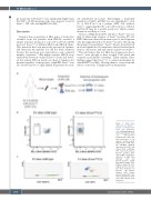
Figure 6. HLA allele expression by Exon1mut - positive hematopoietic stem and progenitor cells. (A) Establishment of induced pluripotent stem cell (iPSC)-derived hematopoietic stem cells from monocytes of an aplastic anemia patient with Exon1mut (UPN 333). (B) HLA-Bw6 (B5401) expression by CD34+ cells derived from a wild-type iPSC clone (left) and an Exon1mut -positive iPSC clone (right). (C) Exon1mut detection in DNA from
wild-type (left) Exon1mut-positive (right) iPSC-derived CD34+ cells. Numbers below the scat- tergram denote the vari- ant allele frequency of Exon1mut. AA: aplastic anemia; UPN: unique patient number; iPS cells: induced pluripotent stem cells; VAF: variant allele frequency.
and
1588
haematologica | 2021; 106(6)
