
Page 73 - 2021_06-Haematologica-web
P. 73
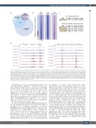
RUNX1-EVI1 blocks RUNX1 and EVI1 driven cell fate
A
BC
Figure 5. RUNX1-EVI1 disrupts RUNX1 binding but also binds to unique binding sites. (A) Venn diagram showing the number of uniquely called and overlapping RUNX1 and RUNX1-EVI1 chromatin immunoprecipitation sequencing (ChIP-seq) peaks. (B) Comparison of RUNX1 binding in -dox and +dox treated cells, RUNX1 ChIP- seq peaks were ranked according to the fold difference of the normalized plus doxycycline (+dox) /-dox tag count across a 2 kilobase (kb) window. The tag-density of the RUNX1-EVI1 ChIP-seq peaks is plotted alongside. The bar alongside indicates the +dox specific sites (blue), shared sites (black) and -dox specific sites (orange). (C) De novo motif enrichment was conducted within the RUNX1-EVI1 ChIP-seq peaks, at both the unique sites and those which were also bound by RUNX1 in either -dox, +dox or both. (D) Genome browser screenshots showing an example site where both RUNX1 and RUNX1-EVI1 bind (left, Ncor2 locus) and where RUNX1-EVI1 binds in the absence of RUNX1 (right, Ccnc locus).
The EHT process is a true cellular transition that is cell cycle independent.26 Whilst we observed a reduction in the proportion of HP related to perturbation of the EHT, we also noted a considerably lower total number of HP than in the control (Figure 2A). This result could be explained by an increase in cells not actively cycling in G0/G1 (48.2% vs. 55.4% without and with RUNX1-EVI1, Figure 2B), specifically by increased numbers of cells in G0 (5.4% with RUNX1-EVI1 compared to <1% without, Figure 2C). We also found a modest increase in apoptotic cells (Online Supplementary Figure S2A).
In order to investigate whether RUNX1-EVI1 expression not only disrupted the EHT, but also affected the ability of progenitor cells to terminally differentiate, we induced RUNX1-EVI1 in newly forming progenitors and placed the progenitors into methylcellulose colony forming unit assays. We first carried out colony-forming unit assays in the absence of dox in the methylcellulose medium. RUNX1-EVI1 protein was quickly lost following the with- drawal of dox (Online Supplementary Figure S2B). Despite RUNX1-EVI1 being absent, we saw an overall reduction in
the number of colonies formed (Figure 2D; Online Supplementary Figure S2C) primarily accounted for by a reduction the number of myeloid (-dox 58±13, +dox 42±11 per 5,000 HP seeded) and erythroid colonies (-dox 26±7, +dox 20±4), with a concomitant increase in the proportion of mixed lineage colonies, or those of unclear lineage (-dox 29±9, +dox 26±6, Figure 2E). The colonies which did form were generally smaller with fewer healthy cells (Figure 2F), which may relate to the previously observed increase in apoptosis (Online Supplementary Figure S2A). When RUNX1-EVI1 expression was either induced or maintained in the HP when they were plated into methylcellulose, colony-forming capacity was further reduced (Figure 2D). Importantly, we did not see enhanced myeloid differentia- tion after RUNX1-EVI1 induction when HP were cultured in liquid or semi-solid medium (Figure 2E; Online Supplementary Figure S2D). These data suggest that whilst RUNX1-EVI1 expression affects the differentiation capaci- ty of HP, there was some reversibility of this phenotype and the continued expression of RUNX1-EVI1 caused ongoing changes.
haematologica | 2021; 106(6)
1575
D
