
Page 213 - 2021_06-Haematologica-web
P. 213
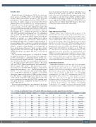
Table 1. Clinical and pathological features of 11 patients with breast implant-associated anaplastic large cell lymphoma.
Hypoxia signature in BIA-ALCL
Introduction
Anaplastic large cell lymphomas (ALCL) are a heteroge- neous group of CD30-positive T-cell lymphomas with varying clinical presentation, prognosis, and molecular pathogenesis.1 Breast implant-associated (BIA)-ALCL is a rare form of ALCL arising in association with breast implants placed for reconstructive or cosmetic purposes.2 It typically occurs in the peri-implant capsule and/or effu- sion an average of 9 years after implant placement. The cytological and immunophenotypic features of BIA-ALCL are similar to those of systemic and primary cutaneous ALK-negative ALCL, including the presence of hallmark cells, CD30 positivity, and frequent loss of T-cell markers such as CD3 and CD5. The prognosis of patients with BIA-ALCL is associated with clinical stage, particularly the presence or absence of a mass-forming lesion and/or locoregional lymph node involvement, which influence the therapeutic approach.3,4 Complete surgical excision of the peri-implant fibrous capsule is essential and sufficient in patients without a mass or lymph node involvement, whereas systemic chemotherapy is recommended in those with advanced disease.3,5 Based on these distinct clinical features, the revised World Health Organization (WHO) classification recognizes BIA-ALCL as a provision- al entity.2
The molecular pathogenesis of BIA-ALCL remains incompletely understood. Recent studies have suggested a possible relationship to underlying chronic allergic reac- tion and bacterial biofilm infection.6,7 Rearrangements of ALK, DUSP22, and TP63 are consistently absent, referred to as the triple-negative (TN) genetic subtype.8,9 Recurrent JAK1 and STAT3 gene mutations have been identified9-12 and, like many other ALCL,13,14 BIA-ALCL shows consis- tent activation of the JAK-STAT3 pathway as detected by immunohistochemistry for Tyr705-phosphorylated STAT3.9,12,15 In vivo studies have demonstrated that inhibi- tion of JAK-STAT signaling by sunitinib or ruxolitinib effectively suppresses growth of TLBR cell lines derived from BIA-ALCL,15,16 suggesting potential therapeutic utility of these drugs for patients with advanced disease. In addi- tion to mutations affecting the JAK-STAT signaling path- way, gene alterations in epigenetic modifiers are also fre- quent in BIA-ALCL.17
However, these findings have not identified a molecular profile of BIA-ALCL that is unique to the peri-implant
microenvironment in which it originates. Identification of unique molecular features specific for BIA-ALCL could lead to discovery of biomarkers: that improve early detec- tion, diagnosis, and follow-up; identify candidate targets for therapy or preventive strategies; and provide justifica- tion to upgrade the WHO classification of BIA-ALCL from a provisional to a definite entity. We therefore interrogat- ed the gene expression profile of BIA-ALCL.
Methods
Gene expression profiling
Human studies were conducted with approval of the Institutional Review Boards at Mayo Clinic and The University of Texas MD Anderson Cancer Center. We performed RNA sequenc- ing on formalin-fixed paraffin-embedded tumor tissue from 11 patients with BIA-ALCL (Table 1). All were female and their mean age was 55 years (range, 44-73 years). All had received textured implants. As described previously,14 RNA from AllPrep extraction was used to prepare sequencing libraries (TruSeq RNA Access, Illumina) and sequenced on a HiSeq 4000 (Illumina). Reads were aligned to hg38 with MAP-RSeq18 modified to use the STAR align- er.19 Gene-level read counts based on Ensembl version 78 were transformed into reads per kilobase per million mapped reads (RPKM). Gene expression data were compared to those of 24 pre- viously sequenced non-BIA-ALCL of TN genetic subtype (10 pri- mary cutaneous ALCL and 14 systemic ALK-negative ALCL14). Gene set enrichment analysis (GSEA) was performed using GSEA software (Broad Institute) as described previously.14
Immunohistochemistry
Immunohistochemistry for CA9 was carried out on formalin- fixed paraffin-embedded sections of 17 BIA-ALCL and 48 non- BIA-ALCL (from patients with a mean age of 54 years). The WHO subtypes of these latter were primary cutaneous ALCL (n=13), ALK-negative ALCL (n=24), and ALK-positive ALCL (n=11). Genetic subtypes included 11 ALK-positive, ten with DUSP22 rearrangements, two with TP63 rearrangements, and 25 TN. Deparaffinized tissue sections were heated in pH 6.0 citric acid buffer in a steam cooker for 30 min. After incubation with 3% hydrogen peroxide for 10 min and 5% bovine serum albumin for 10 min, the slides were incubated with anti-CA9 rabbit monoclon- al antibody (1:100 dilution, clone D47G3; Cell Signaling Technology) at 4°C overnight. Sections were then incubated with horseradish peroxidase-conjugated anti-rabbit secondary antibody
Patient # Age Sex
151 46 F
224 55 F 403 47 F 425 45 F 2680 74 F 2896 65 F 3176 61 F 3177 57 F 3181 76 F 3183 63 F
3184 41 F
ALK DUSP22-R TP63-R T* N* M* Stage* Subtype†
Neg. Neg. Neg. T1 N0
Neg. Neg. Neg. T2 N0
Neg. Neg. Neg. T1 N0
Neg. Neg. Neg. T2 N2
Neg. Neg. Neg. T1 N0
Neg. Neg. Neg. T4 N0
Neg. Neg. Neg. T4 N0
Neg. Neg. Neg. T1 N0
Neg. Neg. Neg. T2 N0
Neg. Neg. Neg. T4 N0
Neg. Neg. Neg. T4 N0
M0 IA Insitu
M0
M0
M0
M0
M0
M0
M0
M0 IB M0 IIA
IB Tumor type IA Insitu
IIB Tumor type IA Insitu
IIA Tumor type IIA Tumor type
IA Insitu Tumor type Tumor type
Ageinyears.F:female;Neg.:negative;R:rearrangement.*TNM(tumor-node-metastasis)stagingaccordingtoClemensetal.49 †HistologicalsubtypeaccordingtoLaurentetal.17
haematologica | 2021; 106(6)
M0 IIA
Tumor type
1715
