
Page 93 - 2021_05-Haematologica-web
P. 93
Zebrafish Hax1-associated neutropenia
ABC
D
EFG
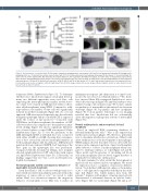
Figure 1. Characterization of zebrafish hax1. (A) Schematic comparison showing syntenic conservation of the hax1 loci in humans and zebrafish. (B) A neighbor-join- ing phylogenetic tree of Hax1 proteins, which was performed with 1,000 bootstrap replications. Am, Astyanax mexicanus; Bt, Bos taurus; Ci, Ciona intestinalis; Dr, Danio rerio; Hs, Homo sapiens; Mm, Mus musculus; Ol, Oryzias latipes; Pt, Pan troglodytes; Rn, Rattus norvegicus; Ss, Salmo salar. (C) Spatial hax1 expression by whole mount in situ hybridization analysis from 5 to 20 hours post-fertilization (hpf). (D) Confocal image of double fluorescent in situ hybridization of hax1 (magenta) and cmyb (green) at 20 hpf. (E-G) Spatial hax1 expression at 24 (E), 48 (F) and 96 (G) hpf. Arrows in C, E, and F indicate hax1 expression in the hematopoietic site. A sense probe was used as a negative control (E, right panel). Note that the images shown in E are two images stitched together. y: yolk; ye: yolk extension. Scale bars: 100 mm (C, E-G), 50 mm (D).
formation (Online Supplementary Figure S3). To determine whether hax1 knockdown impairs neutrophil develop- ment, two different approaches were used. First, cells expressing the neutrophil-specific marker myeloid peroxi- dase (mpo) were stained in MO-injected embryos (here- after called morphants) using WISH. Compared to wild- type embryos, the number of mpo+ cells was significantly reduced in all three morphants (Figure 2B). As a second approach, MO were injected separately into the zebrafish transgenic tg(mpo:gfp) embryos in which GFP is expressed under the control of mpo promoter.30 Consistent with WISH data, in all three morphants, the numbers of GFP+ cells were reduced in comparison to the numbers in unin- jected siblings (Figure 2C) and no significant difference was observed when a control MO was injected (Online Supplementary Figure S4). To test the specificity of the MO, full-length mRNA of zebrafish hax1 was co-injected with either e1-MO or e2-MO, showing that overexpres- sion of hax1 rescued the reduced neutrophil numbers in both morphants (Figure 2D). In addition to MO-mediated gene knockdown, transient CRISPR-Cas9 targeting of the hax1 gene in the tg(mpo:gfp) line was performed. Compared with the non-injected siblings, hax1 crispants showed significantly fewer GFP+ cells in the trunk region at 2 dpf (Online Supplementary Figure S5). Collectively, our findings suggest that hax1 has a role in zebrafish neu- trophil development.
Normal phagocytic activity and migratory behavior of neutrophils in hax1 morphants
Given that Hax1 is able to interact with proteins asso- ciated with cytoskeleton machinery and is involved in the migration of cancer cells in vitro,38 we tested to what extent Hax1 loss-of-function impairs the migratory behavior of neutrophils in vivo. One way to induce an
inflammatory response and chemotaxis is to inject bacte- ria into the notochord of zebrafish embryos.35 We, there- fore, injected Alexa 594-conjugated Staphylococcus aureus debris into this region (Figure 3A) and then embryos were analyzed using confocal microscopy. We found a similar accumulation of neutrophils in the infected site between wild-type (Figure 3B, top panels) and hax1 morphants (Figure 3B, bottom panels). Time-lapse in vivo imaging revealed that hax1 knockdown did not substantially affect the migration or phagocytic activity of neutrophils (Figure 3C).
Normal erythropoiesis and monopoiesis in hax1 morphants
Based on single-cell RNA sequencing databases of zebrafish hematopoietic cells,36,37 hax1 is also expressed in erythrocytes and macrophages. We, therefore, performed tests to determine whether loss-of-function of Hax1 could affect erythropoiesis or the development of macrophages. WISH analysis showed that there were no detectable dif- ferences in the expression patterns of gata1 and hemoglobin alpha embryonic 1.1 (hbae1.1) in the morphants when com- pared to wild-type embryos at 1 dpf (Figure 4A and B). These results and the presence of circulating red blood cells in the morphants (data not shown) suggest that hax1 is dispensable for embryonic erythropoiesis.
We next examined to what extent the development of myeloid cells was affected in hax1 morphants. We tested the expression of pu.1, a regulator of monocytic differen- tiation.39 In 2 dpf hax1 MO-injected embryos, the number of pu.1+ cells appeared comparable with that in the wild- type fish (Figure 4C), which mimics data from CN patients.40 Analysis with markers of mature myeloid cells demonstrated a substantial decrease in l-plastin+ leuko- cytes (Figure 4D), lyz+ neutrophils (Figure 4E) and g-csfr+
haematologica | 2021; 106(5)
1313
