
Page 28 - 2021_05-Haematologica-web
P. 28
D.M. Ross et al.
ducibly detected following removal of the drug. This implies that the accumulated p-JAK could act as a patho- logical signaling node as the drug level falls in patients if drug is abruptly stopped or a dose missed.
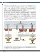
More recently, Tvorogov and co-authors extended these findings to show that accumulation of p-JAK2 is due to resistance of p-JAK2 to ubiquitination and degradation while bound to ruxolitinib.51 Specifically, immunoprecipi- tation with an anti-p-JAK2 antibody produced a band for p-JAK2 in untreated cells, but not in ruxolitinib-treated cells, consistent with an altered conformation induced by the drug. Accordingly, it was noted that cells exhibiting delayed phosphorylation kinetics (i.e., those with wild- type JAK2) were significantly more sensitive to ruxolitinib cytotoxicity and growth inhibition than cells with rapid phosphorylation kinetics (cells with JAK2 V617F). This was an important observation that raised the possibility that ruxolitinib allows drug-bound JAK protein to escape the negative feedback loops of dephosphorylation and degradation (Figure 2B).
To test this, recombinant JAK2 kinase was treated with a phosphatase in the presence of ruxolitinib. Ruxolitinib
blocked the dephosphorylation by PTP1B for up to 20 h whereas reduction of Tyr1007/1008 phosphorylation nor- mally occurred within 2 h.51 In keeping with this, ruxoli- tinib prevented any detectable ubiquitination of JAK2 after cytokine stimulation. These results suggested that binding of ruxolitinib induces a conformational change that con- ceals Tyr1007/1008 from phosphatase access, and ruxoli- tinib-bound p-JAK2 is no longer susceptible to dephospho- rylation or degradation.
Interestingly, primary CALR-mutant cells did not exhibit either JAK2 phosphorylation in the presence of ruxolitinib or striking withdrawal signaling to the same degree as the JAK2 V617F samples. CALR-mutant myelofibrosis cells showed undetectable levels of activated JAK2 Tyr1007/1008 phosphorylation in the presence of ruxoli- tinib and in some CALR+ samples and in a CALR-mutated cell line (MARIMO) total JAK2 protein was difficult to detect, if not completely absent. This is consistent with a number of emerging reports using CALR-mutated models of myelofibrosis55-57 suggesting fundamental differences in the nature of JAK activation in myelofibrosis patients with mutated CALR (Figure 1).
ABC
Figure 2. Signaling in JAK2 V617F cells before, during, and after discontinuation of ruxolitinib. Schematic representation of JAK-STAT activation in JAK2 V617F myelo- proliferative neoplastic cells. (A) JAK2 V617F leads to increased signaling through STAT5/STAT3/ERK leading to proliferation and inflammation. (B) In the presence of a type 1 JAK2 inhibitor, such as ruxolitinib, signaling through the JAK-STAT pathway is abrogated, but so too is ubiquitination and degradation of JAK2, leading to accu- mulation of p-JAK2. Signaling through MEK/ERK remains activated and contributes to disease persistence. (C) Ruxolitinib discontinuation leads to transiently increased signaling through the accumulated pool of p-JAK2 with inflammatory symptoms.
1248
haematologica | 2021; 106(5)
