
Page 26 - 2021_05-Haematologica-web
P. 26
D.M. Ross et al.
resistance is an exciting area of ongoing research that is beyond the scope of this review.
Molecular aspects of CALR and ruxolitinib
Somatic mutations in exon 9 of the gene coding for the endoplasmic chaperone protein calreticulin (CALR) are found in 70-80% of patients with JAK2-negative myelofi- brosis and account for ~30% of myelofibrosis cases over- all.2,27 Virtually all CALR mutations in MPN are small insertions or deletions clustered in exon 9, resulting in a +1 frameshift and loss of the last four amino acids (KDEL) that form the endoplasmic reticulum retention signal, leading to altered distribution of CALR. The two com- monest mutations are a 52 bp deletion (type 1, which is present in 45-53% of patients) and a 5 bp insertion (type 2, present in 32-41% of patients). Although more than 50 CALR mutations have been described, the majority can be classified as type 1-like or type 2-like based on bioinfor- matic predictions of protein structure.28 This functional classification is clinically relevant as it influences progno- sis in myelofibrosis.29,30 The reason for differing prognosis is unclear, but there are biological differences that might be relevant: type 1 mutations eliminate all the negative charge of the C-terminal domain eliminating its calcium binding, thus potentially activating proteases and protein misfolding in the endoplasmic reticulum,31 and potentially altering the chaperone function of CALR.
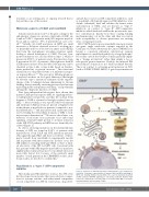
Data from independent laboratories have shown that mutant CALR protein requires MPL for signaling and fac- tor-independent cell growth, and that the normal lectin domain of CALR is essential to bind glycosylated sites on MPL.32-34 More recently, it was reported that both mutant and wild-type CALR proteins are present at higher levels in the plasma of myelofibrosis patients (compared to nor- mal individuals),35,36 and may function in a paracrine fash- ion by binding the extracellular domains of MPL to facil- itate receptor dimerization.34,36 However, the relative con- tribution of autocrine versus paracrine versus endosomal signaling of mutant CALR protein to aberrant activation of the JAK-STAT pathway in MPN has not been fully elu- cidated (Figure 1).
As stated, activating mutations in the juxtamembrane domain of MPL are found in 5-10% of patients with myelofibrosis. Both CALR and MPL mutations presum- ably signal through JAK2, and there is evidence of thera- peutic benefit from ruxolitinib in experimental models and in patients with these mutations. In these cases, the JAK inhibitor is binding wild-type JAK2, so there is limit- ed selectivity of the TKI for MPN cells. Consistent with this, patients with JAK2-negative myelofibrosis may have slightly inferior clinical responses to ruxolitinib (discussed more fully below) but, because of the relatively small number of such patients in the COMFORT studies, addi- tional studies are needed to confirm this.37
Ruxolitinib is a “type I” ATP-competitive inhibitor
Historically, pan-JAK inhibitors (such as AG-490) were first developed as molecules that were substrate-compet- itive for tyrosine residues, and either mixed competitive or non-competitive for ATP. In recent years, drug devel-
opment has focused on ATP-competitive inhibitors, such as ruxolitinib. All clinically approved JAK inhibitors (rux- olitinib, fedratinib) bind and stabilize the kinase-active conformation of JAK2, and are known as “type I” inhibitors, in contrast to “type II” ATP-competitive inhibitors which bind and stabilize the protein in its inac- tive conformation. Exactly how these varying binding mechanisms play out in the clinic and their association with susceptibility to disease persistence are exciting areas of ongoing research.
JAK2 V617F mutations are remarkable among recurrent oncogenic single nucleotide variants reported in the Catalogue of Somatic Mutations In Cancer (COSMIC) for having a consistent inflexible substitution, namely replacement of a small hydrophobic by a large hydropho- bic residue outside the catalytic domain, generally imply- ing a “change in function” rather than simply a loss or non-specific gain in function. Notably, the mutated JH2 pseudokinase domain does not bind ruxolitinib directly. This is in contrast to activating point mutations in FLT3 (such as D835Y, D835H, F691L) or gate-keeper mutations
Figure 1. Activation of JAK-STAT signaling in CALR-mutant cells is inhibited by ruxolitinib. Schematic representation of mutant CALR (mCALR) trafficking in MPN cells showing inhibition by ruxolitinib. The exact mechanism(s) by which mCALR activates JAK-STAT signaling are still being elucidated. Mutant CALR leaves the endoplasmic reticulum and is associated with MPL to promote homodimerization and activation of JAK2.
1246
haematologica | 2021; 106(5)
