
Page 163 - 2021_05-Haematologica-web
P. 163
Mantle cell lymphoma with MYC-R
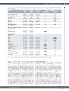
Table 1. Comparison of features of mantle cell lymphoma with MYC rearrangment (MYC-R), mantle cell lymphoma without MYC-R and MYC/BCL2 double-hit lymphoma.
Features
MCL with
MYC-R
(n=27)
63 (47-85)
67% (18/27) 20:7 92% (24/26) 96% (23/24) 33% (4/12) 77% (20/26) 65% (17/26) 40% (10/25) 58% (15/26)
11% (3/27) 89% (24/27) 26% (7/27)
70% (7/10) 31% (4/13) 73% (19/26) 35% (9/26) 50% (4/8) 86% (12/14) 80% (12/15) 69% (9/13) 71% (12/17) 90 (15-100)
67% (16/24)
33% (8/24)
33% (8/24)
MCL with non-MYC-R (n=61)
61.5 (33-85)
54% (33/61) 44:17 92% (55/60) 83%(50/60) 21% (4/19) 68% (39/57) 46% (23/50) 44% (22/50) 44% (22/50)
46% (28/61) 54% (33/61) 26% (16/61)
90% (28/31) 26% (8/31) 87% (53/61) 11% (6/56) 67% (6/9) 97% (28/29) 17% (6/36) 9% (3/33) 65% (13/20) 60 (2-100)
24% (13/54)
74% (40/54)
62% (29/47)
MYC/BCL2
DHL (n=95)
60.5 (33-86)
59% (56/95) 64:31 66% (58/88) 44% (33/75) 13% (7/52) 56% (49/88) 86% (55/64)
85% (60/71)
93% (68/73) 5% (3/63) 96% (87/91) 31% (14/45) 94% (83/88) 85% (39/46) 78% (36/46) 63% (12/19) 85 (20-100)
51% (44/86)
49% (42/86)
32% (27/85)
P value of MCL with MYC-R
Age (years), Median (range)
vs. MCL without MYC-R
0.25
0.21 0.80 1.00 0.17 0.68 0.61 0.22 0.81 0.32
0.004*
0.88
0.14 0.73 0.11 0.01* 0.64 0.22 0.0001* 0.0001* 1.00 0.004*
0.001*
0.03*
vs. MYC/BCL2 DHL
0.21
0.32
1.00
0.01* 0.0001* 0.20 0.11 0.03*
0.0001* 0.0001* 0.0001* 0.42
0.22 0.70 0.46 0.73 0.53
0.25
1.00
Age ≥60 (years)
Sex (Male:Female)
Stage IV
BM-Positve
CNS-Positve
Extranodal Sites ≥2
Elevated LDH (>618 U/L)
Elevated WBC(>11.0 × 10⁶/mL)
High MIPI/ High or High-Intermediate IPI Morphology for MCL
Classic Blastic/Pleomorphic Leukemic Non-Nodal
Immunophenotype
SOX11+
BCL6+
CD5+
CD10+
MUM-1+
BCL2 (≥50%)
MYC (≥40%)
MYC/BCL2 dual-expression P53 (≥20%)
Ki67, Median(range) Treatment
Intensive chemotherapy
Other immuno/chemotherapy
Initial CR
Blank: not available; BM: bone marrow; CNS: central nervous system; CR: complete remission; LDH: lactate dehydrogenase; MIPI: Mantle Cell Lymphoma International Prognostic Index; MCL: mantle cell lymphoma; DHL: double hit lymphoma; intensive chemotherapy (R-CHOP): rituximab, cyclophosphamide, doxorubicin, vincristine, prednisone; other immuno/chemotherapy (R-Hyper-CVAD): rituximab, cyclophosphamide, vincristine, doxorubicin, dexamethasone; WBC: white blood cell; *P<0.05.
with de novo MCL and 13 (48%) patients acquired MYC-R at time of disease progression or transformation from classic to blastoid/pleomorphic MCL. There were 13 (48%) cases diagnosed initially in lymph nodes, 11 (41%) cases in bone marrow and three cases in other tissue sites. Most patients presented with high stage (Ann Arbor stage IV) disease, high frequency of involvement of bone mar- row or other extranodal sites, and elevated white blood cell (WBC) count and serum lactate dehydrogenase (LDH) level (Table 1). The involved extranodal sites included the bone marrow, spleen, central nervous system, gastroin- testinal tract, peripheral blood, pleural fluid, pancreas, chest wall and soft tissue. A leukemic non-nodal form of MCL, defined as MCL with peripheral blood, bone mar- row and sometimes spleen involvement but without sig- nificant lymphadenopathy by WHO classification, was present in seven (26%) patients. Twenty-six patients had available clinical data to calculate the Mantle Cell Lymphoma International Prognostic Index (MIPI) score37 and 15 (58%) patients had a high-risk MIPI score (Table 1).
Pathologic characteristics
Twenty-four (89%) cases of MCL associated with MYC-R cases had blastoid (n=19) (Figure 1) or pleomor- phic (n=5) morphologic features and three cases were clas- sic type. Eleven of 14 (79%) de novo MCL with MYC-R showed blastoid (n=10) or pleomorphic (n=1) morpholo- gy. All 13 patients with MCL that acquired MYC-R during disease progression presented with classic MCL at initial diagnosis, but had blastoid (n=9) or pleomorphic (n=4) morphology at the time of emergence of MYC-R.
All lymphomas were positive for one or more pan-B-cell antigens and were negative for pan-T cell antigens. As expected, all MCL with MYC-R cases expressed cyclin D1 (27 of 27, 100%), and most cases expressed SOX11 (7 of 10, 70%), and MYC (12 of 15, 80%). Concurrent MYC and BCL2 expression was observed in 9 of 13 (69%) MCL cases assessed. Nineteen of 26 (73%) cases were positive for CD5 (one case not assessed); the CD5-negative cases included four de novo MCL, two neoplasms which appar- ently lost CD5 at the time of detection of MYC-R, and one
haematologica | 2021; 106(5)
1383
