
Page 130 - 2021_05-Haematologica-web
P. 130
F. Arruga et al.
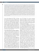
Figure 5. Inhibition of A2A signaling in vivo partially restores immune competence. (A) Flow cytometry analysis of leukemic cells (defined as the CD5+/B220+ cell population) in the spleen, peripheral blood (PB) and bone marrow (BM) of TCL1 mice treated or not with SCH58261 (n=20 mice for each group). (B) In vitro effects of SCH58261 (SCH) on leukemic cell viability. Cells were cultured either alone or in the presence of 10 mM fludarabine (Flud) for 48 h. Where indicated, cells were supplied with 50 mM adenosine (ADO) every 12 h or treated with 10 mM SCH58261, starting from the beginning of the experiment and without replenishing it. ADO confers a survival advantage to TCL1 leukemic cells and protects them from spontaneous and fludarabine-induced apoptosis. SCH58261 efficiently reverts these effects without resulting per se in major apoptosis of leukemic cells, although a slight increase of fludarabine-induced apoptosis was observed in the presence of A2A inhibitor. Results are from 17 independent experiments and statistics were calculated with the Wilcoxon test for paired data. (C) Percentage of regulatory T cells (Treg) out of the total CD4+ cell population progressively increases in TCL1 mice, analyzed at different time-points after injection of leukemic cells, compared to WT not injected mice. In vivo treatment with SCH58261 significantly prevents Treg differentiation. Analyses were performed on 20 TCL1 mice for each condition (different time-points and SCH58261 treatment) compared to seven WT not injected mice. Statistics were calculated using the Mann-Whitney test for unpaired data. (D) Percentage of CD107a+ cells out of CD8+ T lymphocytes in terminally ill vehicle-treated TCL1 mice (n=12) compared to SCH58261-treated mice (n=8) or to WT C57BL/6 (n=12). Statistics were calculated using the Mann-Whitney test for unpaired data, when comparing TCL1 and WT mice, and the Wilcoxon test for paired data, when analyzing, within the same experimental group, the effect of stimulation/inhibition of A2A compared to the untreated condition. (E) Percentage of inter- leukin (IL)-2+ (left panel) and interferon (IFN)-γ+ (right panel) cells out of CD8+ T lymphocytes in terminally ill vehicle-treated TCL1 mice (n=12) compared to SCH58261- treated mice (n=8) or to WT C57BL/6 (n=12). Statistics were calculated using the Mann-Whitney test for unpaired data, when comparing TCL1 and WT mice, and the Wilcoxon test for paired data, when analyzing, within the same experimental group, the effect of stimulation/inhibition of A2A compared to the untreated condi- tion. (F) Characterization of monocyte subpopulations in TCL1 mice, analyzed at different time-points after injection of leukemic cells, compared to WT not injected mice, show a dramatic increase of the patrolling (M2-like) subset at the expense of the inflammatory (M1-like) one. In vivo treatment with SCH58261 significantly rescues skewing of monocytes, restoring the inflammatory subset. Monocytes were defined as the Lin–CD11b+F4/80+ cell fraction and subpopulations were identi- fied on the basis of LY6C and CD43 expression (inflammatory: LY6C+CD43–; intermediate: LY6C+CD43+; patrolling: LY6C–CD43+). Analyses were performed on 20 TCL1 mice for each condition (different time-points and SCH58261 treatment) compared to seven WT not injected mice. Statistics were calculated using the Mann- Whitney test for unpaired data.
response element binding protein (CREB).17,18 For this rea- son, we set-up a phosflow assay to rapidly assess CREB phosphorylation status upon exposure of different cell sub- sets to A2A agonists. The results indicated that leukemic cells, as well as CD4 and CD8 subsets, showed constitu- tively higher levels of CREB phosphorylation, likely indi- cating an activated status of the pathway. Addition of CGS, a specific A2A agonist, further enhanced CREB phos- phorylation, suggesting that in this context inflammatory responses are blunted. As a control, cells were pre-treated with the A2A antagonist SCH58261 before exposure to the agonist, completely inhibiting CREB phosphorylation (Figure 4C, Online Supplementary Figure S2C and D). These data were confirmed on leukemic cells by demonstrating PKA activation, which was higher in leukemic B cells than in normal B cells, further enhanced by the agonist and blocked by the antagonist (Figure 4D).
In vivo targeting of A2A restores immune competence Data so far indicate that progression of leukemia is accompanied by increased hypoxia, which leads to the modulation of the adenosinergic axis, with production of adenosine and activation of its signaling pathway. The hypothesis is that this may lead to progressive adaptation of the immune system to these conditions, with inhibition of potentially tumor-specific T cells. If this is true, inhibi- tion of A2A signaling should prove beneficial to immune
cells, reconstituting their functional properties.
In order to address this issue, we treated leukemic-bear- ing mice with the commercially available SCH58261 A2A antagonist, which was shown to be well tolerated in ani- mals. The A2A antagonist was administered every other day for 2 weeks, starting from 4 days after injection of the leukemic cells. Sixteen hours after the last administration, animals were euthanized and leukemia dissemination as
well as immune cell populations were analyzed.
In line with our previous data showing that extracellular adenosine exerts mild protection from drug-induced apop- tosis on leukemic cells,19 SCH58261 treatment in monotherapy in vivo had minimal effects on tumor bur- den, with no significant differences highlighted between vehicle- or SCH58261-treated mice when looking at the percentage of leukemic cells colonizing the spleen, periph- eral blood or bone marrow (Figure 6B). Similar to what was observed in primary human CLL cells, in vitro expo-
sure of TCL1 leukemic cells to adenosine significantly improved cell viability in the presence of standard chemotherapeutic agents (e.g., fludarabine). Blocking A2A signaling with SCH58261 reverted the cytoprotective effect of adenosine but did not result in a major apoptotic response of TCL1 leukemic cells when used as a single agent. In contrast, it slightly but significantly potentiated the antileukemic effect of fludarabine, opening the way to combination studies (Figure 6A).
The administration of SCH58261 as monotherapy was however very useful for identifying the major contribu- tion of A2A signaling in leukemia progression in vivo. In fact, as expected, leukemia progression was accompanied by marked immune adaptation, as documented by the sig- nificant increase in regulatory T cells (Treg), a finding also in line with what was observed in patients.20 Treatment with SCH58261 prevented accumulation of Treg; the per- centage of Treg out of total CD4+ cells was 15.6% in ter- minally ill vehicle-treated mice versus 11.8% in SCH58261-treated mice (Figure 5A, Online Supplementary Figure S3A). In addition, T cells from leukemic mice were dysfunctional, showing reduced expression of the degran- ulation marker CD107a (Figure 5B, Online Supplementary Figure S3B), an effect again reverted upon A2A blockade. T cells from terminally ill mice also secreted significantly less interleukin-2 and interferon-γ compared to cells obtained from WT animals, both at baseline and after short-term incubation with phorbol 12-myristate 13- acetate (PMA) and ionomycin (Figure 5C, Online Supplementary Figure S3B), again in line with data from patients.21 Compared to the T cells from vehicle-treated TCL1 mice, those from mice given SCH58261 showed restored ability to secrete interleukin-2 and interferon-γ, both at baseline and following maximal cellular activation with PMA and ionomycin (Figure 5C, Online Supplementary Figure S3B). Lastly, treatment with SCH58261 had a notable impact on the myeloid subset of cells. Indeed, leukemia progression was marked by an increase in patrolling monocytes at the expense of inflam- matory ones, suggesting an imbalance similar to that observed in patients.22 Importantly, inhibition of A2A sig- naling reverted this adaptation, restoring the inflammato- ry component and limiting the patrolling one (Figure 5D, Online Supplementary Figure S3C).
The results suggest that targeting adenosine signaling
1350
haematologica | 2021; 106(5)
