
Page 95 - 2021_04-Haematologica-web
P. 95
Tumor suppressor activity of RXR in AML
low nM concentrations of retinoids and exhibited speci- ficity between RARA and RXRA ligands (Online Supplementary Figure S2D-F). Although the RXRA LBD is used in this assay, the ligand-binding pocket is highly con- served between RXRA, RXRB, and RXRG, and no RXR subtype-specific compounds have yet been identified, suggesting that natural ligands that activate RXRA are
likely to cross-react with RXRB and RXRG.25 We noted the presence of mCherry+GFP+ cells in both BM and spleen from multiple mice transplanted using three different pri- mary MLL-AF9 leukemias and engrafted into a total of five different recipients (Figure 1B and E). As a negative control, we transduced UAS-GFP x MLL-AF9 leukemia cells with a retrovirus expressing Gal4-RXRA-DAF2,
AB
CDE
FG
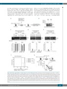
Figure 2. Retinoid X receptors (RXR) act as tumor suppressors in mouse MLL-AF9 leukemias. (A) Schema for leukemia transplant procedure. Rxraflox/flox x Rxrbflox/flox x Mx-Cre bone marrow (BM) cells were collected from the donor mice and transduced as indicated and injected into sublethally irradiated recipient mice. Upon leukemia engraftment, the proportion of Rxra and Rxrb deletion was assessed by polymerase chain reaction (PCR). (B) PCR results from representative donor BM cells with “Flox” indicating the retained and “∆” the deleted allele. (C-E) PCR analysis of Rxra and Rxrb alleles from leukemias that emerged from individual mice. Bar graphs display the quantified percentage of deleted alleles in each unique leukemia. ImageJ software was used for quantification analysis. (F) Kaplan-Meier sur- vival curve of mice injected with Rxraflox/flox x Rxrbflox/flox x Mx-Cre MLL-AF9 leukemic cells (RXR-KO) or Rxraflox/flox x Rxrbflox/flox MLL-AF9 leukemic cells (RXR-flox). Each cohort consisted of five mice. Indicated cohorts were treated with three doses of pIpC on days 5-10 (solid lines). (G) Schema for BM transplant procedure of Lox- stop-Lox-YFP x Mx-Cre mice and YFP evaluation of the hematopoietic cell. Kit+ BM cells from Lox-stop-Lox-YFP x Mx-Cre donor mice were harvested (n=3 donor mice) and transduced with MSCV-MLL-AF9 retrovirus, then injected into sublethally irradiated recipient mice (n=7 recipient mice). The percentage of yellow fluorescent protein (YFP) was quantified by flow cytometry in BM cells before transduction and after leukemia emerged. Each line represents results from an individual recipient mouse.
haematologica | 2021; 106(4)
1011
