
Page 38 - 2021_04-Haematologica-web
P. 38
C.A. Di Buduo et al.
graduated scaffold was meant to support megakaryocyte function by a bone marrow-like structure. To modulate the pore size of the collagen scaffold, a two-stage freezing technique was used, which created a variety of pore sizes with larger pores in the top layer and small pores at the bottom. Based on differential pore sizes this scaffold had a sieving capacity that enhanced the purity of the platelet output. Indeed, the bioreactor was conceived as a twin chamber culture system, whereby one side of the chamber allowed seeding of the cultured hiPSC-derived megakaryo- cytes into the scaffold, while cross-flow-generated shear forces were able to induce platelet release. In these condi- tions, platelet production was improved significantly com- pared to that in 2D cultures. Using a different approach, involving soft gel lithography, Kotha and colleagues were able to use collagen hydrogels as a scaffold to create a microvascular network.103 Endothelial cells were seeded in the microvascular network to form endothelial vessels with a lumen, while megakaryocytes were encapsulated directly in the type I collagen hydrogel. Megakaryocytes were able to migrate to the microvascular network and to extend proplatelets. This bioengineered device provided a tool to study the vascular-megakaryocyte interface during thrombopoiesis. However, one limitation of this model was the size of the vessel (around 100 mm), which was big- ger than sinusoid vessels.
The search for biomaterials that can be chemically and mechanically tailored to entrap bioactive molecules, such as growth factors and extracellular matrix components, while retaining bioavailability, has spawned research into the use of silk fibroin as scaffolds. Silk fibroin from
Bombyx mori silkworm cocoons is a strong but elastic pro- tein that is biocompatible, having low immunogenicity and low thrombogenicity.104,105 Our group designed a silk tube functionalized with components of the extracellular matrix that support platelet production, such as fibronectin, type IV collagen and laminin, and stromal- derived factor-1α, surrounded by a type I collagen hydro- gel and Matrigel or by a silk sponge.105 The structure of the silk sponge was closer to the medullar topography and enhanced adhesion and migration of cells within the structure.105 In the vascular compartment, the presence of stromal-derived factor-1α directed the migration of mature megakaryocytes towards the silk tube. Platelets collected with flow passing through the tube were func- tional and still alive 4 days after collection. It was also shown that co-culture with a monolayer of endothelial cells or functionalization of the silk tube with vascular endothelial growth factor and vascular cell adhesion mol- ecule 1 increases the yield of platelets. This model could be easily adapted to study mechanisms of normal and pathological megakaryopoiesis and for drug screening.69,105
In an attempt to scale-up platelet production for transfu- sions, multi-porous silk sponges have been investigated.106 These sponges were cultured within new modular flow chambers with flow passing through the different pores and megakaryocytes in direct contact with the flow. This enhanced the capacity of platelet production, because of the larger volume of perfusion that allowed an increase of the concentration of cells in the sponge and because of the softer environment functionalized with extracellular matrix components that support proplatelet formation.
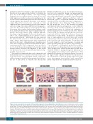
Figure 4. In vivo versus in vitro: an overview of different culture approaches for generating platelets and erythrocytes. In vivo cell maturation occurs in a complex environment in which cells experience different mechanical and biochemical cues due to cell-to-cell and cell-to matrix interactions. In the classical in vitro two-dimen- sional culture, cell contacts, confinement and environmental biomechanics are lost; moreover cells in contact with the plastic are artificially polarized. In the three- dimensional culture, topography and stiffness can be modeled to mimic the native environment. Only cell cultured in flow conditions can recapitulate blood hydro- dynamics. Microfluidic devices have the ability to enable extension of proplatelets and the release of functional platelets. Three-dimensional bioreactors combine the advantages of a three-dimensional environment with flow through the scaffold: mature cells can migrate toward the perfused compartment to release either mature erythrocytes or platelets. Cell culture in agitated or stirred-tank bioreactors has been exploited to allow large-scale production of platelets or erythrocytes. 2D: two- dimensional; 3D: three-dimensional. The figure was created using Servier Medical Art templates licensed under a Creative Commons Attribution 3.0 Unported License (https://smart.servier.com).
954
haematologica | 2021; 106(4)
