
Page 280 - 2021_04-Haematologica-web
P. 280
Letters to the Editor
A
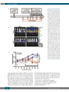
Figure 3. In vivo activity of Bi38-3 in the MM.1Sluc xenograft mouse model. (A) Treatment schedule. Six- to 12-week-old NOD/SCID/IL-2Rγnull mice were inoculated with 5x10⁶ MM.1SLuc cells by tail vein injection (i.v) at day 0, followed, 13 days later, byinfusionof5x106 purifiedhumanT cells, isolated from healthy donors, with (or without) Bi38-3 at a dose of 0.1 mg/kg (blue arrow). Treatment was initiated at day 13 when similar levels of luciferase-expressing MM cells were detected in all mice. Tail vein injections of Bi38-3 (0.1 mg/Kg) or phosphate-buffered saline (PBS, for controls) were repeated daily for 9 days (black arrows). Bioluminescence was measured with the IVIS Imaging System on days 7, 11, 13, 15, 18 and 21 (or 22) after tumor injection (red arrows). (B) Serial bioluminescence imaging was performed to assess myeloma progression/regression. Radiance was measured on the entire body of mice. Images on the left show bioluminescence at 7 days after inoculation with MM.1S myelo- ma cells and before the beginning of the treatment. Images on the right indicate bioluminescence 18 days after inoculation with MM.1S cells and 4 days after treatment with Bi38- 3 (lower panel) or with vehicle (upper panel). The radiance color scale is represented on the right. (C) Longitudinal radiance levels of mice treated with vehicle (blue lines) or Bi38-3 (red lines). Red and blue curves represent groups of nine and 11 mice inoculated with MM.1SLuc and T cells and then treated with Bi38-3 (0.1 mg/kg) and PBS, respec- tively. Black filled triangles indicate the first injection of Bi38-3 or PBS with T cells and white filled triangles indicate Bi-38 or PBS injections every day for 9 days. These experiments were performed with T cells isolated from two independent donors. The normality of populations was estab- lished with the Shapiro-Wilk normality test, and P-values were calculated based on an unpaired Student t test (*P<0.05; **P<0.01; ***P<0.001).
B
C
We report here the development of Bi38-3, a new anti- CD38/CD3 BiTE, which triggers T-cell-mediated lysis of CD38+ MM cells in vitro, ex vivo and in vivo. Interestingly, Bi38-3 provokes no toxicity on B, T and NK cells in vitro and is, therefore, likely to have less "off tumor" effects than AMG424, a recently developed anti-CD38 bispecific antibody.12 Importantly, Bi38-3 efficiently triggers killing of MM cells from patients who are resistant to standard treatments. Furthermore, because it recognizes a specific epitope on CD38 and is devoid of the Fc region, Bi38-3 is expected to be efficient also in relapsed patients follow-
ing daratumumab therapy. Collectively, the data present- ed in this study identifies Bi38-3 as a selective and effi- cient compound for the treatment of MM, which could be used as a front-line agent or at relapse (alone or in combination with other drugs), and which should be evaluated further in MM patients.
Maxime Fayon,1 Carolina Martinez-Cingolani,1 Audrey Abecassis,1 Nathalie Roders,1 Elisabeth Nelson,1 Caroline Choisy,1 Alexis Talbot,1,2 Armand Bensussan,1 Jean-Paul Fermand,1,2 Bertrand Arnulf1,2
and Jean-Christophe Bories1
1196
haematologica | 2021; 106(4)
