
Page 278 - 2021_04-Haematologica-web
P. 278
Letters to the Editor
AD
BE
C
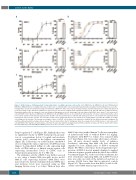
Figure 1. Bi38-3 induces CD38-dependent T-cell-mediated lysis of multiple myeloma cells in vitro. (A-C) KMS11-luc (A), MM.1S-luc (B) and CD38-deficient MM.1S-luc (MM.1S-KO-luc) (C) multiple myeloma cell lines were co-cultured with T cells, isolated from peripheral blood samples from healthy donors, at an effec- tor:target (E:T) cell ratio of 5:1 with increasing concentrations of Bi38-3 for 24 h. Curves represent target cell lysis, monitored by luciferase activity and expressed as the percentage of the untreated condition (0% lysis). Data are the means of independent experiments with four (A), nine (B) and four (C) different donors. Standard deviations (SD) are shown for each concentration. Histograms on the left show target cell lysis induced by Bi38-3 alone, donor T cells alone and T cells with Bi38-3 (101 ng/mL) on KMS11-luc (A), MM.1S-luc (B) and MM1.S-KO-luc (C). (D and E) Fresh tumor plasma cells were collected from buffy coat of bone marrow aspirates from myeloma patients, then CD138+ cells were purified and co-cultured with autologous CD3+ T cells isolated from peripheral blood mononuclear cells at an E:T cell ratio of 5:1 for 24 h. Cultures were analyzed by fluorescence-activated cell sorting (FACS) to monitor the number of CD138+ cells falling into the live gate. The average percentages of lysis of CD138+ cells (relative to the untreated condition) in four different patients at diagnosis (D) and three different patients at relapse (E) are shown. The error bars indicate the SD. Histograms show the average effects of Bi38-3 alone, T cells alone and T cells with Bi38-3 (102 ng/mL) on tumor plasma cells from the same four patients at diagnosis (D) and three patients at relapse (E). SD are shown and P values were determined by a two-sided Mann–Whitney U-test (*P<0.05; **P<0.01; ***P<0.001).
Foxp3+ regulatory T cells (Figure 2D). Similarly, there was no significant toxicity on CD34+ hematopoietic progeni- tors at concentrations below 10 ng/mL and moderate toxicity (>40% survival) at the highest concentrations (Figure 2E). Collectively, our results indicate that Bi38-3 does not impair the surface expression of CD38 and only triggers T-cell-mediated killing of cells expressing high levels of CD38 with no or limited toxicity against cells expressing intermediate levels of CD38, such as hematopoietic progenitors, B, T or NK cells.
The antitumor activity of Bi38-3 was further assessed in vivo using a human MM xenograft mouse model. MM.1S cells expressing luciferase (MM.1Sluc) were injected into the tail vein of immunodeficient (NSG) mice and luciferase levels were measured using an IVIS Imaging System every 3 or 4 days. Thirteen days after
MM.1S injection, purified human T cells were transplant- ed intravenously with or without Bi38-3 (0.1 mg/kg). Treatments with Bi38-3 or vehicle were repeated daily for 9 days (Figure 3A). Seven days after tumor cell injec- tion, all mice showed similar levels of radiance (luciferase), indicating that MM cells had effectively engrafted in host animals prior to Bi38-3 treatment (Figure 3B). While control mice showed rapid tumor pro- gression, all Bi38-3-treated animals displayed a marked reduction in tumor growth within the first 5 days of Bi38- 3 treatment (Figure 3C). At the end of the treatments, the level of luciferase-expressing MM cells in Bi38-3-treated mice was only one tenth of the initial level and was on average 30-fold lower than that in untreated controls (Figure 3C). These results show that Bi38-3 is able to effi- ciently control MM tumor progression in vivo.
1194
haematologica | 2021; 106(4)
