
Page 154 - 2021_04-Haematologica-web
P. 154
J.P. Loftus et al.
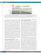
Figure 1. Constitutive SYK signaling occurs in infant acute lymphoblastic leukemia (ALL). Simple Western analysis of splenic lysates from human ALL patient-derived xenograft (PDX) models demonstrated high basal phosphorylated SYK (pSYK) levels in the majority of infant non-KMT2A-rearranged (R) (light blue) and KMT2A-R (dark blue) ALL specimens. pSYK levels were lower in most childhood non-KMT2A-R ALL specimens (red) and absent in splenic tissue from non-leukemia-injected NSG mice (gray). Total SYK levels were similar across all models. ALL PDX model names are specified above corresponding Simple Western data.
We then assessed the ability of ENTO to inhibit leukemia proliferation in vivo in ALL3103 and NH011 (Ph- like ALL with NUP214-ABL1 fusion) PDX mice. ENTO 0.03% and 0.07% chow concentrations administered for 28 days both potently decreased human CD45+ CD19+ ALL cell counts in peripheral blood measured weekly by quantitative flow cytometry and in end-study spleens (Figure 2B and C and Online Supplementary Figure S7). Terminal PK evaluation of ENTO in the periphery con- firmed that high levels of ENTO could be achieved by continuous chow administration (Figure 2D) without sta- tistical difference between the 0.03% and 0.07% treat- ment groups. Simple Western analysis of highly leukemia- engrafted splenic lysates from individual ENTO-treated mice demonstrated marked inhibition of pSYK Y323, cMYC and pERK T202/Y204 as compared to control chow-treated animals after 4 weeks of treatment (Figure 2E) and high correlation between ENTO levels and pSYK and pERK inhibition in well-engrafted ALL3103 PDX mice treated in pharmacodynamic studies for 72 hours with entospletinib (Online Supplementary Figure S8). These results confirmed the on-target inhibition of pSYK and key downstream signaling phosphoproteins by ENTO, sug- gesting that an achieved dose level of 3330-7900 nM in vivo was sufficient to inhibit constitutive pSYK signaling and decrease in vivo leukemia proliferation in an aggressive relapsed infant KMT2A-R ALL PDX model.
In vitro pharmacodynamic inhibition of signaling proteins in infant KMT2A-R models
To extend our observation of ALL cell SYK dependency for proliferation and survival in other KMT2A-R fusion types, we evaluated ENTO in another aggressive multi- ply-relapsed infant ALL PDX model with KMT2A-MLLT1 fusion (ALL135MR) in short-term in vitro cultures and observed dose-dependent inhibition of pERK1/2, pAKTS473, pSTAT5, and cMYC (Figure 3A). Interestingly, similar in vitro incubation of leukemia cells from an infant ALL PDX model with KMT2A-AFF1 fusion and concomi- tant NRASG12D mutation (ALL142MR) with ENTO showed little to no inhibition of the same key pathways (Figure 3B). These data suggest differential signaling effects poten- tially related to specific KMT2A fusion partner and/or RAS-mutant status.
Evaluation of expression signatures in KMT2A-R acute lymphoblastic leukemia subtypes
KMT2A-R ALL has been shown to have distinct gene expression signatures that define B-cell developmental arrest at either the pro-B- and pre-B-cell stages.22 Understanding the signaling pathway dependencies of dif- ferent KMT2A-R fusion proteins in infant ALL cells may lead to more effective therapeutic targeting strategies for this high-risk patient population. To assess potential dif- ferential gene expression signatures, we evaluated the transcription factors HOXA9 and MEIS1, which are known downstream targets of KMT2A. As hypothesized, HOXA9 and MEIS1 expression levels correlated with both KMT2A-R fusion status and specific gene partner (Figure 4A). Infant ALL specimens with KMT2A-MLLT3 and KMT2A-MLLT1 fusions expressed both high HOXA9 and MEIS1, while KMT2A-AFF1 models had high MEIS1 and normal HOXA9 expression. Conversely, infant non- KMT2A-R samples had normal expression levels of HOXA9 and MEIS1. These distinct expression signatures exhibited amongst KMT2A-R samples with different fusion partners are concordant with reports of differential chromatin binding of KMT2A-R fusion proteins leading to distinct gene expression profiles and potentially differen- tial clinical outcomes.1,21
Given the observed stratification of HOXA9 and MEIS1 expression signatures among the KMT2A subgroups, we next assessed protein expression signatures in these sam- ples to evaluate potential correlation. Simple Western analysis of splenic lysates from KMT2A-R and non- KMT2A-R ALL PDX models (Figure 4B) demonstrated that leukemias with different KMT2A fusion partners induced different patterns of signaling activation. High levels of cMYC were detected only in KMT2A-AFF1 models, while KMT2A-MLLT1 models had high SRC, absent PTEN, and high pAKT levels. Regulation of both SRC and PI3K path- ways are known to be potentially SYK-dependent, concor- dant with data from our in vitro studies in ENTO-treated ALL135MR cells (Figure 3A). Overall, differential gene expression signatures between KMT2A-R and non- KMT2A-R ALL subtypes (Online Supplementary Figure S9A) and differences between gene and protein expression sig- natures among the KMT2A fusion subtypes (Figure 4B and Online Supplementary Figure S9B) showed unique signaling
1070
haematologica | 2021; 106(4)
