
Page 115 - 2021_04-Haematologica-web
P. 115
BET and FLT3 inhibition for FLT3-ITD AML
A
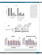
Figure 6. Primary samples co-cul- tured on stroma using quantitative- polymerase chain reaction (qPCR) and immunoblotting. (A) MYC expression in primary acute myeloid leukemia (AML) blasts by qPCR. Primary blasts from a patient with relapsed FLT3-ITD AML were co-cul- tured on stroma and treated under the specified condition. RNA was isolated from cells at 6-hour (h) and 24-h time points and analyzed as described in the Methods section. (B) MYC protein expression in pri- mary AML blasts by immunoblot- ting. A primary FLT3-ITD AML sam- ple (AML2, from Figure 4B) was treated with drug for 3 h, lysed, and probed for MYC as described in the Methods section. β-actin was used as control for protein loading. Protein expression levels were quantified by densitometry.
B
A
B
Figure 7. Colony forming cell assays. Colony assays were performed using normal bone marrow progenitor cells from healthy donors. Cells were cultured either with (A) PLX51107 alone or (B) with 50 nM quizartinib and increasing concentrations of PLX51107 for 10-14 days prior to assessment by microscopy. Averages of three individual experiments (three different donors) are shown; standard error of mean is shown as error bars. BFU: burst-forming unit; CFU: colony-forming unit.
haematologica | 2021; 106(4)
1031
