
Page 178 - 2021_03-Haematologica-web
P. 178
F. Denorme et al.
reperfusion.33 Since VWF is a known modulator of inflam- mation,20 a thromboinflammatory role for VWF in the ischemic stroke brain was proposed. Yet, the inflammato- ry mechanisms by which VWF mediates reperfusion injury in the ischemic stroke brain remain poorly under- stood. Using flow cytometric analysis of single-cell sus- pensions prepared from brain tissue from VWF KO and WT mice and histology experiments, we found that the acute immune response in the brain after stroke was
greatly reduced in the absence of VWF. These results fur- ther corroborate the notion that VWF contributes to cere- bral inflammation in stroke.34-36 In agreement with Khan et al., we found that neutrophil recruitment to the ischemic stroke brain was significantly reduced in VWF-deficient mice compared to that in WT mice.35 Interestingly, we additionally identified a two-fold decrease in the recruit- ment of inflammatory monocytes and a four-fold decrease in the recruitment of T cells to the ischemic
AB
C
D
EF
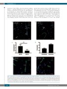
Figure 3. Immunofluorescent visualization of neutrophils and T cells in the ipsilateral hemisphere of mice 24 hours after ischemic stroke brain injury. Transient focal cerebral ischemia was induced in von Willebrand factor (VWF) wild-type (WT) or knockout (KO) mice by occluding the right middle cerebral artery for 60 min. This was followed by 23 h of reperfusion, after which, brain sections were stained for blood vessels and neutrophils or T cells. (A, B) Neutrophils were stained with a marker for Ly6G (red) and blood vessels with a lectin stain (green). Neutrophils are marked with an *. (C) Quantification of the number of neutrophils/mm2 in the infarct core of WT and VWF KO mice (n=3). Data are the mean ± standard deviation. (D) The number of neutrophils within and outside the vasculature (n=3). Data are the mean ± standard deviation. (E, F) T cells were stained with a marker for CD3 (red) and blood vessels with a lectin stain (green). T cells are marked with an *. Scale bars are 20 mm. Images are representative of three mice per genotype.
824
haematologica | 2021; 106(3)
