Page 177 - 2021_03-Haematologica-web
P. 177
VWF-mediated thromboinflammation in stroke
VWF A1 also led to a two-fold reduction of immune cell recruitment in the brains of treated mice compared to that in control mice (7095 ± 2550 vs. 16310 ± 3980, respectively; P<0.005) (Figure 5A). Both myeloid (5195 ± 2259 vs. 11978 ± 3322; P<0.05) (Figure 5B) and lymphoid WBC counts (1529 ± 269 vs. 3017 ± 514; P<0.05) (Figure 5C) were reduced in the ipsilateral hemispheres of VWF A1 nanobody-treated mice. Specifically, inhibition of the VWF A1 domain reduced the number of infiltrated inflammatory monocytes (3726 ± 1824 vs. 8266 ± 2651; P<0.05) (Figure 5D), neutrophils (304 ± 94 vs. 1557 ± 317; P<0.0005) (Figure 5E) and T cells (487 ± 93 vs. 1111 ± 248; P<0.05) (Figure 5F) in the ipsilateral hemisphere of the brain, 24 h after stroke. These data suggest that the inflammatory effect of VWF in ischemic stroke is mediat- ed through the VWF A1 domain.
Discussion
As the main finding from this study, we report that VWF mediates an inflammatory response during cerebral ischemia/reperfusion via the recruitment of inflammatory monocytes, neutrophils and T cells, through a mechanism
AB
that is dependent on the VWF A1 domain. Blocking the A1 domain reduced the inflammatory response and improved stroke outcome.
Current ischemic stroke treatment is aimed at achiev- ing reperfusion of the ischemic brain as soon as possible. When thrombolysis or thrombectomy is initiated, an unsalvageable infarct core often already exists. Reperfusion therapy is therefore aimed at salvaging the penumbra to limit further ischemic brain damage. Unfortunately, rescue of the perfused salvageable penum- bra is not always achieved, which has led to the concept of cerebral ischemia/reperfusion injury.31,32 In recent years, VWF has emerged as an important mediator of cerebral ischemia/reperfusion injury.30 We and others reported that absence of VWF protected mice from ischemic stroke brain injury, without increasing the risk of cerebral bleed- ing.13,14 Intriguingly, the detrimental role of VWF in the ischemic brain appears to be distinct from its role in hemostasis. Whereas hemostatic thrombus formation requires both platelet adhesion and platelet aggregation, the latter does not seem to play a major role in reperfu- sion injury after ischemic stroke.15,16 Besides thrombotic events, ischemic stroke is also characterized by a strong inflammatory response that occurs in the brain after
CD
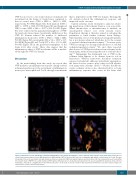
Figure 2. Immunofluorescent visualization of thromboinflammation in the ipsilateral hemisphere of mice 24 hours after ischemic stroke brain injury. Transient focal cerebral ischemia was induced in von Willebrand factor (VWF) wild-type (WT) or knockout (KO) mice by occluding the right middle cerebral artery for 60 min. This was followed by 23 hours of reperfusion, after which brain sections were stained for VWF (red), platelets (green) and nuclei (blue). (A) Only a few platelets were found within the ischemic brain of VWF KO mice. (B-D) Clumps of VWF together with platelets were frequently found attached to the vessel wall within the ipsilateral, stroke-affected hemisphere. Panel (D) is a magnification of the white box in panel C. Scale bars are 50 mm except for that in panel (D) in which the scale bar is 25 mm. Images are representative of three animals per genotype.
haematologica | 2021; 106(3)
823


