
Page 174 - 2021_03-Haematologica-web
P. 174
F. Denorme et al.
VWF is a large multimeric glycoprotein that recruits platelets at sites of vascular injury. Several case-control studies demonstrated increased VWF levels in ischemic stroke patients6-11 and high plasma levels of VWF were found to be an independent risk factor for ischemic stroke.12 Preclinical experiments have shown that mice lacking VWF show significantly reduced brain injury and better functional outcome in experimental models of cere- bral ischemia/reperfusion injury.13,14 Remarkably, initial VWF-mediated platelet adhesion rather than subsequent platelet aggregation contributes to ischemic brain injury with a prominent role for the interaction between the
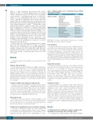
Table 1. Antibody cocktails used to discriminate between different white blood cell subtypes.
Antibody cocktail 1
Antibody cocktail 2
Antigen
CD45 APC-Cy7
CD11b PE-Cy7
Ly6G BV510
Ly6C FITC
CD16/CD32 (Fc-block) Live/Death Fixable Violet Dead Cell Stain Kit
CD45-APC-Cy7
CD11b PE-Cy7
CD11c PerCp-Cy5
CD3e FITC
CD16/CD32 (Fc-block) Live/Death Fixable Violet Dead Cell Stain Kit
Supplier
Biolegend eBioscience Biolegend Biolegend eBioscience
ThermoFisher
Biolegend eBioscience eBioscience eBioscience eBioscience
ThermoFisher
15-19 VWF A1 domain and platelet glycoprotein (GP)Iba. It is
currently not known exactly how platelets and VWF con- tribute to stroke progression in a way that is not strictly related to platelet-thrombus formation. Most likely, VWF- mediated acute inflammation also aggravates acute ischemic stroke brain injury,20 but the exact mechanisms of VWF-mediated inflammatory responses in stroke remain poorly understood.
In this study, we used flow cytometry and a unique nanobody that blocks the VWF A1 domain to investigate the precise role of VWF in the acute cerebral inflammatory response during stroke. We specifically found that neu- trophils, monocytes and T cells were recruited to the brain after stroke through a mechanism that involves the VWF A1 domain.
Methods
A detailed description of the methods can be found in the Online Supplement.
Animals and nanobodies
For this study, 10-week old VWF knockout (KO)21 and littermate wild-type (WT) C57BL/6 mice were used. All animal experiments were approved by the local ethical committee (P050/2017 KU Leuven, Leuven, Belgium) and were performed following the ARRIVE guidelines (www.nc3rs.org.uk), including randomization of treatment and analysis blind to the treatment. Mice were treated with a well-characterized nanobody targeting the VWF A1 domain (KB-VWF-006 bv; 10 mg/kg) or a control nanobody (KB-VWF-004 bv; 10 mg/kg).22
Cerebral ischemia and reperfusion injury model
Transient occlusion of the middle cerebral artery was performed as described previously.17 Briefly, a standardized silicon rubber- coated 6.0 nylon monofilament (6021; Doccol Corp, Redlands, CA, USA) was inserted via the right internal carotid artery to occlude the origin of the right middle cerebral artery. The suture was left in situ for 60 min. Immediately after the start of reperfu- sion, nanobodies were administered intravenously.
Neurological tests
Twenty-four hours after induction of transient middle cerebral artery occlusion, mice were subjected to the modified Bederson test23 and the grip test24 to assess global neurological and motor function, respectively, as described previously.25
Cerebral lesion quantification and assessment of bleeding
To measure cerebral infarct volumes, mice were euthanized 24 h after induction of transient middle cerebral artery occlusion. Coronal brain sections (2 mm thick) were stained with 2% 2,3,5- triphenyl-tetrazolium chloride (TTC, Sigma-Aldrich, St Louis,
MO, USA) to visualize cerebral infarcts. The presence of cerebral hemorrhages was assessed macroscopically.
Flow cytometry
Twenty-four hours after stroke, mice were euthanized and per- fused with 20 mL of phosphate-buffered saline. Single-cell suspen- sions were made of the ischemic (ipsilateral) and non-ischemic (contralateral) hemispheres as previously described.26 Next, cells were incubated with appropriate antibody cocktails (Table 1) con- taining Fc-blocker. Live cells were stained with Live/Death violet cell viability staining (L34963; ThermoFisher; Waltham, MA, USA) after which they were fixed and analyzed with a FACSVerse flow cytometer (BD, Franklin Lakes, NJ, USA) and BD Facs Suite soft- ware.
Immunofluorescence
Twenty-four hours after stroke, mice were euthanized, and the brains were dissected. Nine micron thick sections were stained for the presence of neutrophils (rat anti-mouse Ly6G, 1/500, eBioscience, San Diego, CA, USA), T cells (Armenian hamster anti- mouse CD3e, 1/500, Biolegend, San Diego, CA, USA), VWF (rab- bit anti-human VWF, 1/1500, Dako, Santa Clara, CA, USA) or platelets (rat anti-mouse GPIX, 1/100, emfret, Würzburg, Germany). A lectin dye (FITC conjugated lectin from Lycopersicon esculentum, 1/500, Sigma-Aldrich) was used to stain the microvas- culature.
Statistical analysis
Statistical analysis was performed with Graph Pad Prism Version 8.1.2. Prior to statistical analysis, a D’Agostino and Pearson normality test was used to check data distribution. One-way analysis of variance with a Dunnett post-hoc test or a Mann- Whitney test was used for statistical comparison of infarct size and immune cell infiltration when applicable. In the case of non-para- metric data (Bederson and grip-test scores) a Kruskal-Wallis test with post-hoc Dunn correction was performed. Infarct size is rep- resented as mean ± standard deviation. Bederson and grip-test scores are shown as scatter plots with the median. Immune cell recruitment is shown as a minimum-maximum box plot, with the median.
Results
von Willebrand factor deficiency reduces immune cell recruitment to the brain after ischemic stroke
To examine the cerebral immune cell response mediat-
820
haematologica | 2021; 106(3)
