
Page 39 - 2021_02-Haematologica-web
P. 39
Coagulation factor VII, hemostasis and thrombosis
FVII zymogen circulates in plasma at low concentration (500 ng/mL, 5nM) and the extracellular proteolytic cleav- age1 of a minute portion (about 1%, 0.1 nM) of FVII giving rise to FVIIa, occurs between residues Arg152-Ile153, (Figure 2)2 producing the light and heavy chains. The origin of FVIIa in plasma is still debated. Substantially decreased levels of plasma FVIIa in individuals with congenital FIX deficiency suggest that the generation of FVIIa is depend- ent on an activation product of FIX. Recently, it has been proposed that different forms of activated FIX (FIXα and FIXβ) participate in a reciprocal activation mechanisms of FVII(a) and FIX(a) (white and red-curved arrows, Figure 3, left panel) that would not require a protein cofactor.24
The FVIIa heavy chain contains the domain character- ized by the (chymotrypsin) serine protease family catalytic triad. The light chain contains the calcium binding, vitamin K-dependent domain (gamma-carboxyl-glutamic, GLA) and two epidermal growth factor-like domains, essential for the interaction with membranes and other proteins2 of the coagulation cascade.
Considerable variation in FVIIa plasma concentrations between individuals has been reported (Figure 4),25 exceed- ing one order of magnitude. Differently from other serine protease of the coagulation cascade, FVIIa displays a plas- ma half‐life (2-3 hours) remarkably close to that of the FVII zymogen, which might be explained by the “zymogen- like” properties of FVIIa that have been weakened by mutagenesis, thus increasing its activity.26
FVIIa interacts with TF, a membrane receptor exposed following vascular lesion (extrinsic pathway). The FVIIa- TF complex,1 essentially conserved in all jawed vertebrates,27 activates both FIX and factor X (FX)28 (Figure
3) on the platelet surface in the presence of Ca2+, leading to the generation of thrombin and the subsequent deposition of fibrin.
The crystal structure of the complex between the active- site-inhibited FVIIa and the cleaved, soluble extracellular domain of TF (sTF) revealed the details of the contoured embrace of FVIIa and sTF domains and the extensive inter- molecular contacts.29 Neutron and X-ray scattering experi- ments30 suggested that FVIIa in solution has an elongated domain structure with significant flexibility, which allows rapid interaction with sTF over a large surface area to form the high-affinity complex (dissociation constant, KD 2−5 nM).
The FVIIa-TF complex is highly dependent on specific lipids for physiological activity.2 Activated phospholipid membranes host both TF, an integral membrane protein, and FVIIa, which binds membranes through its GLA domain. Externalization of phosphatidylserine to the outer membrane leaflet and allosteric TF disulphide bond exchanges (decryption on membranes)4 make TF the FVIIa- activating cofactor. The FVIIa-TF complex formation shapes the FVIIa catalytic site by allosteric interactions and increases its activity up to 106–fold. As a matter of fact, cir- culating FVIIa is the active portion of the total FVII mass only after high-affinity binding with TF. These FVIIa-TF complex-specific molecular events that provide FVIIa with its physiological activity are believed to represent the true initiation of the extrinsic pathway (Table 1).
The physiological negative control of the FVIIa-TF com- plex (Figure 3) occurs through the reversible inhibition by tissue factor pathway inhibitor (TFPI),31 mediated by the TFPI Kunitz domain 1 and with protein S as cofactor.32 An
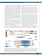
Figure 2. Schematic diagram of the F7 gene and factor VII protein expression. Upper part: Exons are numbered (1-9) and colored in accordance with the encoded protein domains (lower part). Exon 2 is in parenthesis because it is not included in the most abundant mature mRNA. FVII: factor VII; F7: factor VII gene; KB: kilobase. In addition to the complete nuclear transcript the most represented FVII mRNA is indicated. Lower part: Protein domains are indicated with different colors. GLA, tri- angle, γ-carboxyglutamic acid-containing domain. EGF, trapezium, epidermal growth factor-like domain. Green box, promoter. The complete intracellular protein, and the circulating forms are depicted. The numbers of residues in the pre-pro-leader sequence (n=60), in the circulating forms (n=406) and in the light (n=152) and heavy (n=254) chains are indicated. The interchain disulphide bridge is also depicted (yellow bracket).
haematologica | 2021; 106(2)
353
