
Page 328 - 2021_02-Haematologica-web
P. 328
Case Reports
AB
CD
EF
G
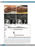
Figure 1. Clinical, brain magnetic resonance imaging (MRI) and chest computed tomography (CT) images of tubercolosis (TB) and fungal infections, and schematic summary of treatments. (A) Localized abscesses on lower limbs at the sites of polyethylene glycol-modified adenosine deaminase injection before surgical incision. (B) Localized abscesses on lower limbs at day +100 after the third haplo-HSCT. Surgical incision was performed before HSC-gene therapy (GT). (C) Brain MRI at diagnosis of intracerebral TB granuloma. Sagittal and coronal post-contrast T1W brain MRI images show a hypothalamic contrast enhancing lesion suggestive for tuberculous granuloma. (D) Brain MRI at day +100 after the third haplo-HSCT showing marked reduction of the tuberculous granuloma. (E) CT images of the lungs at time of aspergillosis diagnosis after the second haplo-HSCT. Axial chest CT images with lung window (left) and mediastinal window (right) show a left lower lobe pulmonary mass compatible with pulmonary aspergillosis. (F) CT images of the lungs at day +100 after the third haplo-HSCT show- ing marked improvement. (G) Schematic representation of the treatment given before and during the third haplo-HSCT to control secondary HLH and prevent graft failure. MPD: methylprednisolone; VP-16: etoposide; CTX: cyclophosphamide; ATG: anti-thymocyte globulin; Cs-A: cyclosporine-A.
642
haematologica | 2021; 106(2)
