
Page 306 - 2021_02-Haematologica-web
P. 306
Letters to the Editor
AB
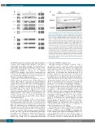
Figure 2. The SMAD1 promoter region is hypermethylated in Hodgkin lym- phoma cell lines and patients’ samples. (A) Methylation analysis by bisul- fite sequencing of regions A1(1), A4(3) and B2(5) within the SMAD1 pro-
C moter in a panel of Hodgkin lymphoma (HL) cell lines. Each circle repre- sents one CG dinucleotide; black circles indicate methylated, white circles indicate unmethylated cytosines. Each line represents one clone. Two or three clones were sequenced per sample. X indicates aligned mismatches between genomic and bisulfite sequences. The cell line KMH-2 has a known frameshift mutation at the SMAD1 locus. (B) SMAD1 expression at the protein level after no treatment (-) or treatment with 1 mM decitabine (+) for 96 h of two HL cell lines as determined by western blotting. Results from two independent experiments are exemplified. Decitabine-treated samples of the DEV cell line are not shown because of lack of protein after demethylating treatment. (C) Methylation analysis by bisulfite sequencing of regions A1(1), A4(3) and B2(5) within the SMAD1 promoter in samples from three patients with classic HL. Each circle represents one CG dinu- cleotide; black circles indicate methylated, white circles indicate unmethy- lated cytosines. Each line represents one clone. Two or three clones were sequenced per sample. X indicates aligned mismatches between genomic
and bisulfite sequences.
the SMAD1 promoter and yielded four hypermethylated cell lines (L428, KMH-2, DEV, HDLM-2), that differed substantially, particularly regarding their methylation of region A4(3), from cell lines showing no evidence of pro- moter hypermethylation (L1236, L540) (Figure 2A). Importantly, KMH-2 is known to be SMAD1 p.ADTP220fs mutant (https://portals.broadinstitute.org/ ccle/page?cell_line=KMH2_haematopoietic_and_lymphoid_tis sue), which additionally points towards a potential role of this gene silencing in lymphomagenesis.
The impact of the promoter methylation status of SMAD1 on protein expression was further addressed by western blot analysis, comparing one hypermethylated cell line (DEV) with a cell line without hypermethylation (L1236). Concordantly, the expression of SMAD1 dif- fered clearly, without detectable protein in the DEV cell line (Figure 2B). When treated with the DNA methyl- transferase inhibitor decitabine, which reverses, among others, the hypermethylation of SMAD1 promoters, the SMAD1-negative cell line DEV died immediately after exposure. As expected, the expression of SMAD1 was not affected by treatment in the L1236 cell line, which is not hypermethylated.
To obtain further clinical evidence, the promoter methylation status of SMAD1 was assessed in samples from three patients with classic HL. To do this, we used our newly developed, flow sorting-assisted technique for HRS cell enrichment from formalin-fixed and paraffin- embedded tissues, allowing for targeted genetic analysis of DNA isolated from classic HL tumor cells.9 In this col- lective, not only the A4(3) promoter region of SMAD1, but also the A1(1) region was substantially hypermethy- lated (Figure 2C). Furthermore, SMAD1 promoter hyper- methylation was identified in sorted tumor-infiltrating plasma cells, fitting with the immunohistochemically
noted lack of SMAD1 in plasma cells.
Although risk-adjusted standard treatment of HL is
successful in over 90% of patients, relapses after salvage therapy and refractory cases represent oncological chal- lenges and run an unfavorable clinical course with limited therapeutic options. In this context, demethylating agents such as decitabine and azacytidine may be of potential therapeutic interest and have already shown promising effects. At clinically relevant concentrations, decitabine has been documented to inhibit the growth of classic HL cell lines in vitro and a single case observation of regressing relapsed classic HL as an unexpected “side effect” of azacytidine has been reported in a patient suf- fering from concomitant myelodysplastic syndrome.10,11 Decitabine and azacytidine inhibit DNA methyltrans- ferases, and thereby reverse promoter hypermethylation of SMAD1.12 Importantly, promising results with decitabine were also obtained in DLBCL cell lines lacking SMAD1 expression due to promoter hypermethylation.5 Four days of treatment were sufficient to restore SMAD1 transcription and protein expression in a subset of initial- ly SMAD1-negative DLBCL cell lines, an effect corrobo- rated by observations in a patient-derived xenograft DLBCL mouse model with proven SMAD1 promoter hypermethylation.5 Analogous to this, we treated the SMAD1-negative HL cell line DEV with decitabine. The cells died immediately after exposure, possibly due to marked responsiveness to decitabine.
Since therapeutic reversion of SMAD1 promoter hyper- methylation would be of potential relevance only in the presence of TGF-β receptors (TGFBR), we analyzed pub- lically available (Gene Expression Omnibus [GEO] acces- sion n. GSE12453) and our own (GEO accession n. GSE147387) gene expression data from primary HRS and LP cells and cell lines to estimate whether HRS and LP
620
haematologica | 2021; 106(2)
