
Page 305 - 2021_02-Haematologica-web
P. 305
Letters to the Editor
SMAD1 promoter hypermethylation and lack of SMAD1 expression in Hodgkin lymphoma: a potential target for hypomethylating drug therapy
Hodgkin lymphoma (HL) is an immunologically active lymphoid neoplasm composed of a few (usually 1-10%) neoplastic Hodgkin and Reed-Sternberg (HRS) cells or lymphocyte-predominant (LP) cells and >90% non-neo- plastic cells, mainly T- and B-lymphocytes, plasma cells, macrophages, eosinophils and fibroblasts. The substan- tial amount of reactive cells in HL is supposed to be the net effect of a complex signaling network of cytokines and chemokines secreted by either the HRS cells or non- neoplastic cells.1 One component of this network is transforming growth factor beta (TGF-β), which is pro- duced by HRS cells and cancer-associated fibroblasts. TGF-β unfolds its immunosuppressive impact by stimu- lating tumor-infiltrating T-lymphocytes (TIL) to differen- tiate into anergic, tumor-promoting, regulatory T cells (Treg).2 Additionally, TGF-β inhibits natural killer cells - one of the key components of the innate anticancer immunity.3 Interestingly and still poorly understood, the HRS cells themselves seem to remain unaffected by the tumor-suppressive properties of TGF-β.4
Recent studies on diffuse large B-cell lymphoma
(DLBCL) revealed a previously unknown tumor-suppres-
5
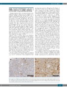
specimens from patients suffering from all subtypes of classic HL (77 nodular sclerosis [NS]; 48 mixed cellularity [MC]; 7 lymphocyte-rich [LR]; 5 lymphocyte-depleted; and 6 unclassifiable classic HL) and 14 routine samples from patients suffering from nodular lymphocyte-pre- dominant HL (NLPHL). We analyzed all these instances for immunohistochemical expression of SMAD1. Importantly, to guarantee retained antigenicity, only cases containing (physiologically) SMAD1-positive endothelia were considered. We found that all NLPHL (14/14 cases; 100%) and the great majority of classic HL (138/143 cases; 97%) displayed SMAD1-negative LP and HRS cells, respectively (Figure 1A and B). Single HRS cells stained faintly for SMAD1 in five cases only (2 NS; 2 MC; and 1 LR classic HL). With respect to non-neoplastic cells, 65/143 classic HL (45%) showed moderate (15-49% of TIL) up to abundant (≥50% of TIL) amounts of SMAD1 positive surrounding TIL, thus being potentially suscepti- ble to the suppressive influence of TGF-β (Figure 1A and B); in NLPHL, 11/14 (79%) cases displayed abundant SMAD1-expressing TIL, including TIL involved in roset- ting around LP cells (Online Supplementary Figure S1). The presence of abundant SMAD1-expressing TIL did not correlate with disease stage, patients’ age, gender, pres- ence of B symptoms, association with Epstein-Barr virus (EBV) or outcome, while showing significant correlations with the NS subtype (45/77 NS cases, i.e. 58%, compared
sive signaling axis involving SMAD1 as a downstream
to 20/66 non-NS cases, i.e. 30%, P=0.025 χ
2 test) and
messenger of TGF-β. SMAD1 functions as an intracellu- lar signal transducer between extracellular TGF-β and the nucleus, where it modulates the transcription of target genes. This signaling cascade was shown to be recurrent- ly inactivated in DLBCL, mainly by hypermethylation of five promoter regions surrounding the SMAD1 transcrip- tion start site, which finally generates a significant growth advantage for lymphoma cells.5 In the course of these investigations, we noted that SMAD1 was not expressed in HRS cells of screened HL cases. This led us to hypothesize that the absence of SMAD1 expression in HRS cells may mechanistically be linked to their resist- ance to the tumor-suppressive effects of TGF-β.4
with the amount of FOXP3-positive Treg (Rho=0.351, P=0.000053 Spearman correlation), which both, in turn, may be directly linked to the effects of TGF-β, promoting sclerosis and a shift towards Treg differentiation.2 In con- trast, surrounding plasma cells seemed to lack SMAD1 expression, potentially rendering them insensitive to the pro-apoptotic and anti-proliferative signals of TGF-β.7 With regard to plasma cells, this largely fits with the newly described negative prognostic impact of their increased numbers in classic HL.8
To strengthen our hypothesis, we investigated the pro- moter methylation status of the SMAD1 gene in six dif- ferent HL cell lines, including one NLPHL cell line (DEV) exactly as described elsewhere.5 Methylation analysis by bisulfite sequencing was successful for three regions of
In order to further elucidate this finding, we analyzed 132 well-characterized archival tissue-microarrayed cases,6 and 11 conventional routine lymphadenectomy
AB
Figure 1. Expression of SMAD1 in classic Hodgkin lymphoma. (A) Tissue microarrayed archival mixed cellularity classic Hodgkin lymphoma with moderate num- bers of SMAD1-positive tumor-infiltrating lymphocytes (TIL) and a few strongly staining endothelia. Note that all Hodgkin and Reed-Sternberg (HRS) cells are neg- ative. (B) Diagnostic lymphadenectomy of a nodular sclerosis cHL with abundant SMAD1-positive TIL and a few strongly staining endothelia. Note that all HRS cells are negative. Immunoperoxidase staining, original magnification 400x.
haematologica | 2021; 106(2)
619
