
Page 244 - 2021_02-Haematologica-web
P. 244
M.M. Ouseph et al.
Loss of chromosome Y in ≥75% metaphases
is significantly associated with a morphological diagnosis of a myeloid neoplasm, especially myelodysplastic syndrome
A WHO-defined diagnosis was made in 39/73 patients (53% of the total): 35 patients were diagnosed with differ- ent subtypes of MDS (MDS with single lineage dysplasia, MDS with multilineage dysplasia, MDS with ring siderob- lasts and multilineage dysplasia, MDS with excess blasts- 1, MDS with excess blasts-2, and therapy-related MDS), as shown in Table 1. Four patients were diagnosed with myelodysplastic/myeloproliferative neoplasm with ring sideroblasts and thrombocytosis. No other MDS/MPN subtypes were identified and none of the patients met cri- teria for MPN or AML.
Among the remaining 34 patients who did not meet diagnostic criteria for any WHO-defined myeloid neo- plasm, 14 had minimal dysplasia not meeting the 10% diagnostic threshold for a diagnosis of MDS and one patient had a hypocellular bone marrow suggestive of aplastic anemia. The remaining 19 patients had a morpho- logically normal bone marrow evaluation. Of note, 73% of patients with minimal bone marrow dysplasia not diag- nostic of MDS had blood cytopenias at presentation, while only 30% of patients with a morphologically nor- mal bone marrow study had peripheral blood cytopenias; however, this difference did not reach statistical signifi- cance due to small numbers (P=0.32; Wilcoxon/Kruskal- Wallis test, χ2 approximation).
There was a significant association between higher per- centages of metaphases with LOY and diagnosis of myeloid neoplasia (Figure 1A). Bone marrow samples with ≥75% LOY had a very high likelihood of a morpho- logical diagnosis of a myeloid neoplasm (P=0.004; Fisher exact test, Pearson χ2 P-value), while samples with <25% LOY were associated with no/minimal dysplasia (P=0.0484; Fisher exact test, Pearson χ2 P-value).
Loss of chromosome Y in ≥75% metaphases is significantly associated with progression to myelodysplastic syndrome
Subsequent bone marrow evaluation had been per- formed on 34 patients who did not meet criteria for a WHO-defined diagnosis of myeloid neoplasia at the initial presentation with LOY, with an interval of 0.4 to 152.8 months (median 34.1 months) after the initial bone mar- row evaluation. In the subsequent marrow evaluation, 4/7 patients (57%) with ≥75% LOY had a marrow morpho- logical diagnosis of MDS, compared to only 1/27 patients with <75% LOY (P<0.0001; log-rank test, χ2 = 22.979) (Figure 1B).
Loss of chromosome Y in ≥75% metaphases
is significantly associated with mutations in myeloid neoplasia-related genes
Among the 73 LOY patients with NGS performed at presentation, 25/32 (78%) patients with ≥75% LOY had pathogenic mutations in tested myeloid neoplasia-associ- ated genes. The frequency of mutations was associated with LOY burden: 5/8 (63%) patients with 50-74% LOY, 5/10 (50%) with 25-49% LOY, and 8/23 (35%) with <25% LOY had mutations (Table 2, Online Supplementary Tables S2 and S3). The mean number of mutations per patient was 2.2 in samples with ≥75% LOY, 1.3 in sam- ples with 50-74% LOY, 0.8 in samples with 25-49% LOY,
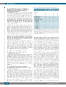
Table 1. Age and pathological diagnoses in the study population. [%] LOY
25-49% 50-74% ≥75% 25-49 1002
Age (years)
50-74 75-100
Pathological diagnosis Nodysplasia
Minimal dysplasia, not diagnostic of MDS MDS/t-MDS
MDS-SLD MDS-MLD MDS-RS-MLD MDS-EB-1 MDS-EB-2 t-MDS
MDS/MPN-RS-T
11 4 11 6
8 4 6 3
9 3 6 1 2 2 0 0 0 0 1 0 0 0 0 0
411 419
4 4 14
320 1 4 110 0 3 0 1 0 1 1 1 0 4
<25%
[%] LOY: percentage of metaphases with loss of Y chromosome; MDS: myelodysplastic syndrome; MDS-SLD: MDS with single lineage dysplasia; MDS-MLD: MDS with multilin- eage dysplasia; MDS-RS-MLD: MDS with ring sideroblasts and multilineage dysplasia; MDS-EB-1: MDS with excess blasts-1; MDS-EB-2: MDS with excess blasts-2; t-MDS: thera- py-related MDS; MDS/MPN-RS-T: myelodysplastic/myeloproliferative neoplasm with ring sideroblasts and thrombocytosis.
and 0.6 in samples with <25% LOY. Bivariate analyses demonstrated that ≥75% LOY was significantly associat- ed with greater likelihood of having somatic mutations commonly associated with myeloid neoplasm (P=0.0009; one-way ANOVA) and a higher number of such muta- tions (P=0.0002; one-way ANOVA) (Figure 2). Multivariate analysis demonstrated a positive correlation between total number of mutations as well as number of mutated genes and % LOY (row-wise method) (Figure 3, Online Supplementary Tables S4 and S5). In multivariate analysis using a logistic regression model with effective likelihood ratio test, % LOY was found to be a statistical- ly significant predictor of diagnosis of myeloid neoplasia (odds ratio [OR] 1.03, 95% confidence interval [95% CI]: 1.02-1.04; P=0.0005) per 20% increase in LOY; OR 6.17 (95% CI: 2.15-17.68; P=0.0007) for ≥75% LOY) (Table 3).
The most commonly mutated genes were TET2, SF3B1, U2AF1, ZRSR2 and ASXL1 (Table 2, Online Supplementary Tables S2 and 3). Analysis of variance demonstrated that the variant allele frequencies (VAF) for pathogenic variants were significantly higher in patients with ≥75% LOY than in those with <25% LOY (P=0.000027, F-ratio=9.4; one-way ANOVA) (Table 2). Patients with ≥75% LOY had an overall higher incidence (50%) of mutations in spliceosome components (SFB31, SRSF2 and U2AF1), JAK2 and RUNX1 compared to the incidence of 9% in patients with LOY <25%, 10% in those with 25-49% LOY and 13% in patients with 50- 74% LOY (P=0.011; Pearson χ2 test) (Table 2).32 In addi- tion, patients with ≥75% LOY had a higher incidence (50%) of two or more mutations compared to the inci- dence of 9% of in patients with <25% LOY, 10% in those with 25-49% LOY and 38% in patients with 50-74% LOY (P=0.01; Pearson χ2 test) (Table 2). Furthermore, a large subset of patients with ≥75% LOY had these muta- tions in combination with mutations in TET2, DNMT3A and/or ASXL1 (28%), compared to 4% of those with <25% LOY, 10% of 25-49% LOY patients and 25% of 50-74% LOY patients (P=0.01; Pearson χ2 test) (Table 2).
558
haematologica | 2021; 106(2)
