
Page 197 - 2021_02-Haematologica-web
P. 197
ATM-induced mitophagy in MCL
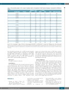
Table 1. Flow cytometry analysis of the response of primary mantle cell lymphoma to FCCP-induced mitophagy is independent of ATM kinase
activity.
Mitophagy Status Dy m
Status
1 t(11;14)
2 t(11;14)
3 t(11;14)
4 t(11;14)
5
6 t(11;14)
7 t(11;14)
8
9 t(11;14) 10
11 t(11;14)
12 t(11;14)
13 t(11;14)
14 t(11;14)
15 t(11;14)
16 t(11;14)
17 t(11;14)
18 t(11;14)
19 t(11;14)
20 t(11;14)
21 t(11;14)
mtDNA copy number
524
580 718 1621 ND 405 ND 489
t(11;14) status
IR status
-
- + - - + - + + + + - + + + + - - + + +
DMSO FCCP
352 358
420 260 290 200 256 120 252 200 250 200 152 155 321 322 244 230 703 287 346 280 235 235 280 350 365 505
DMSO FCCP
90 45
120 36 40 20 32 18 177 82 88 57 55 51 58 32 260 180 54 16 175 50 170 77 195 40 336 99
mROS
20
153 5970 28 377 20 297 23 830 27 640 ND 2711 ND 236 ND 573 14 468 210 512 60 1215 150 1141 125 1151 18 372 55 ND
17 20 28
84 50 29 21
552 312 575 620 360 400 622 632 1120 660 1022 963
265 40 188 58 50 28 198 63 70 53 305 162
Livecells(5x10⁶)wereirradiated(5Gray,asinFigure1G).CellswerestainedwithPE-ATMSer1981 andFITC-γH2AXSer139 todeterminetheirATMkinasestatus(OnlineSupplementary
), or APC-Mitotracker deep red to determine mitochondria mass and deduce their mitophagy response.Untreated cells were stained with MitoSOX Red to determine the basal level of mitochondrial reactive oxygen species. Mitochondrial DNA copy number was analyzed from total cellular DNA isolated from untreated cells by quantitative polymerase chain reaction analysis (see Methods). ND: not
Figure S8) or treated with FCCP (75 mM for 3 h) and stained with PE-TMRE to determine mitochondrial membrane potential (DΨ determined.
m
first described molecular role of ATM in mitochondrial autophagy. Pharmacological targeting of mitophagy through ATM and Parkin, in combination with other drugs or stresses, may offer opportunities to control tumor progression because of the acute sensitivity of tumor cells to mitochondrial dysfunction.
Disclosures
No conflicts of interests to disclose.
Contributions
A.S. conceptualized the study, designed experiments, generat- ed and analyzed data, and wrote the manuscript. CMS con- tributed and provided critical reagents related to autophagy studies and was involved in designing and analyzing the protein half-life and mRNA qPCR studies. HVV contributed in Seahorse XF96 Analyzer and in OCR studies. M.A. performed NTP assays and HPLC analysis. B.A.K. and J.P contributed in qPCR analysis of mtDNA experiments. KB and A.S. con- tributed in FCS assays related to cytotoxicity. JKB and WNH contributed in confocal analyses. TKP contributed critical
reagents and technical help related to ATM kinase function. SSN identified patients and provided primary B-cell lymphoma cells and their cytogenetic characteristics. VG supervised the research project, analyzed data, obtained funding, and reviewed the manuscript. TKP, WNH, CMS and VG contributed in writ- ing/editing the manuscript.
Acknowledgments
We thank Dr. Sankar Mitra, Methodist Research Institute, Houston, TX, USA for crtically reviewing the manuscript. We also thank Dr. Richard Youle, NIH for providing us with the YFP-Parkin plasmid, Dr. Noriyuki Matsuda, Tokyo Metropolitan Institute of Medical Science, Tokyo, Japan for pro- viding us with the GFP-Parkin construct and Rakesh Sharma, Department of Lymphoma/Myeloma, MD Anderson Cancer Center, Houston, TX, USA for processing and distributing the primary B-cell lymphomas. This work was supported by a CLL Global Research Foundation Alliance grant. A part of this work was performed in the Flow Cytometry and Cellular Imaging Facility, supported in part by the NIH through MD Anderson’s Cancer Center Support grant CA016672.
References
1. Brandon M, Baldi P, Wallace DC. Mitochondrial mutations in cancer. Oncogene. 2006;25(34):4647-4662.
2. Fulda S, Galluzzi L, Kroemer G. Targeting
mitochondria for cancer therapy. Nat Rev
Drug Discov. 2010;9(6):447-464.
3. Galluzzi L, Joza N, Tasdemir E, et al. No
death without life: vital functions of apop- totic effectors. Cell Death Differ. 200815(7): 1113-1123.
4. Sabharwal SS, Schumacker PT.
Mitochondrial ROS in cancer: initiators, amplifiers or an Achilles' heel? Nature Rev Cancer. 2014;14(11):709-721.
5. Vander Heiden MG, Cantley LC, et al. Understanding the Warburg effect: the metabolic requirements of cell proliferation. Science. 2009;324(5930):1029-1033.
haematologica | 2021; 106(2)
511
