
Page 175 - 2021_02-Haematologica-web
P. 175
PAI-1 blockade eliminates leukemia stem cells
MT1-MMP, which has been shown to control the motility of HSPC,17–19 in CML-LSC derived from PAI-1 inhibitor treated mice. Mice were transplanted with normal FL LSK cells that were transduced with retrovirus carrying the human BCR/ABL-ires-GFP and were treated with a PAI-1 inhibitor for 7 consecutive days starting at day 7 post transplantation (Figure 5A). As expected, treatment with a PAI-1 inhibitor reduced the expression of iPAI-1 in the LSK fraction of BCR/ABL-GFP+ CML cells, which represent CML-LSC, and at the same time, increased the expression of MT1-MMP in the same fraction of cells (Figure 5B). Furthermore, iPAI-1 blockade altered the expression levels of CD44 and VLA-4, key adhesion/de-adhesion molecules known to determine the localization of hematopoietic cells within the BM14,30,32–34 (Figure 5B). Consistent with this in vivo observation, expression of CD44 and VLA-4 were altered in iPAI-1 OE and KO CML cells, confirming the link between the expression of adhesion/de-adhesion molecules and the iPAI-1 activity (Online Supplementary Figure S1). In order to confirm that iPAI-1 regulates the motility of CML-LSC through the action of MT1-MMP, we examined the directional motility of CML-LSC toward a chemokine gradient using a trans-reconstituted basement membrane (MatrigelTM) migration assay system. Lin–c-Kit+ immature CML cells isolated from PAI-1 inhibitor treated CML mice exhibited significantly higher trans-Matrigel migration activity compared to those of saline-treated mice (Figure 5C). Importantly, addition of anti-MT1-MMP neu- tralizing antibody to the culture abo-lished the enhanced migration activity of immature CML cells (Figure 5C), con- firming the critical role of MT1-MMP in regulating the motility of CML-LSC. We therefore examined the effect of PAI-1 blockage in the localization of CML-LCS in vivo. BM sections were stained with antibodies against hematopoi-
etic lineage markers (CD3, B220, Mac-1, Gr-1, Ter119, CD41, and CD48) and c-kit to identify CML-LSC and were simultaneously stained with antibody against TGF-β to identify niche cells (Figure 5D, the fluorescence images in lower magnification are shown in the Online Supplementary Figure S2), and the sections were evaluated to determine the positional relationships between CML-LSC and niche cells. In PAI-1 inhibitor-treated mice, BCR/ABL-GFP+Lin–c- kit+ CML-LSC were frequently found further away from TGF-β-expressing niche cells (Figure 5D), in contrast to vehicle-treated mice in which CML-LSC were often in contact with, or within close proximity to, TGF- -express- ing niche cells (Figure 5D). Importantly, the administration of an anti-MT1-MMP neutralizing antibody counteracted the observed detachment of CML-LSC from niche cells in PAI-1 inhibitor-treated mice (Figure 5D). These results sug- gest that iPAI-1 activity is an important determinant of CML-LSC’ sensitivity to TKI, whereby controlling the MT1-MMP dependent retention of CML-LCS in the BM protective environment.
Blockade of iPAI-1 activity in combination with TKI efficiently eliminates CML-LSC
As shown earlier in this study that CML-LSC exhibited higher iPAI-1 expression (Figure 1), we focused on the effectiveness of iPAI-1 targeting therapy on primitive CML-LSC. Recipient mice that received BCR/ABL-GFP+ cells obtained from the BM of mice transplanted with human BCR/ABL-ires-GFP retrovirus transduced FL LSK cells were treated with IM alone or with IM plus a PAI-1 inhibitor for 14 consecutive days (Figure 6A). IM treat- ment alone reduced the percentage of BCR/ABL-GFP+ LSK cells in the BM approximately by half and mildly decreased the spleen size (Figure 6B-C). Notably, the com-
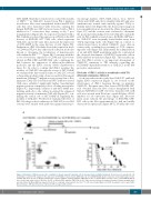
A
BC
Figure 4. Blockade of iPAI-1 increases the sensitivity of chronic myeloid leukemia cells to tyrosine kinase inhibitor treatment. (A) Schema for experiments. (B) Quantitative real-time PCR analysis of the expression of BCR/ABL+ in bone marrow (BM) LSK cells of SCLtTAxBCR/ABL Tg mice that were treated with saline (n=10), imatinib (IM) alone (n=11), or IM plus plasminogen activator inhibitor-1 (PAI-1) inhibitor (TM5614) (n=12). (C) Kaplan Meier survival curves of SCLtTAxBCR/ABL Tg mice treated with saline (n=10), IM alone (n=10), or IM plus PAI-1 inhibitor (TM5614) (n=10). Data represent means ± standard deviation. Statistical significance was determined by Mann-Whitney unpaired t-test (B) or a log-rank non-parametric test (C). P<0.001, by a Kruskal-Wallis test. BCR: breakpoint cluster region; ABL: Abelson kinase: GFP: green fluorescent protein; LSK: Lineage (Lin)Sca1c-Kit.
haematologica | 2021; 106(2)
489
