
Page 127 - 2020_11-Haematologica-web
P. 127
In vivo role and mechanism of platelet migfilin
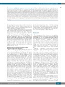
Figure 4 (previous page). Migfilin deficiency reduces outside-in signaling in platelets. (A) Spreading of WT and migfilin-/- platelets on immobilized fibrinogen in the presence or absence of migfilin peptides (5 mM). Images are representative of three independent experiments with similar results. Original magnification 100x. Scale bars, 10 mm (left panel). Quantification of the areas of spread (mm2) of WT and migfilin-/- platelets (mean ± standard error of the mean [SEM], ***P<0.001, ns: no significant difference, Student t test) (right panel). (B) Representative curves and quantification of Mn2+ (0.5 mM)-induced aggregation of washed WT and migfilin-/- platelets in the presence or absence of migfilin peptide. Platelets were stimulated in the cuvettes of a Chrono-log lumiaggregometer in the presence of fibrinogen (25 mg/mL) and under stirring at 1,200 rpm. Experiments were repeated at least four times and the results are shown as mean ± SEM (*P<0.05, ns: no significant difference, paired Student t test). (C) Platelets from WT or migfilin-/- mice were re-suspended with human platelet-poor plasma at a concentration of 4x10⁸/mL, and recombined plasma was stimulated to coagulate with thrombin (0.4 U/mL), then photographed at different time points. Experiments were repeated at least three times. (D) Measurement of early aIIbβ3 outside-in signaling in WT and migfilin-/- platelets spread on fibrinogen or stimulated with Mn2+ (0.5 mM) in the presence of fibrinogen (25 mg/mL) in suspension. At indicated time points, platelets were lysed and analyzed by Western blotting with antibodies recognizing phos- phorylated β3 Tyr747, phosphorylated β3 Tyr759, phosphorylated SFK Tyr416, phosphorylated Syk Tyr525/526, Beta3, Src, and Syk. Experiments were repeated at least three times. (E) WT and migfilin-/- platelets were spread on fibrinogen for 60 minutes (min) or stimulated with Mn2+ (0.5 mM) in the presence of fibrinogen (25 mg/mL) for 10 min, in the presence or absence of migfilin peptides (5 mM). Platelets were lysed and analyzed by Western blotting with antibodies recognizing phosphorylated phosphorylated β3 Tyr747, phosphorylated β3 Tyr759, phosphorylated SFK Tyr416, phosphorylated Syk Tyr525/526, Beta3, Src and Syk. Experiments were repeated at least three times.
the spreading results, neither the rate of clot retraction nor the final volumes of clots differed between WT and migfilin-/- platelets (Figure 4C), indicating an intact late out- side-in signaling upon migfilin deficiency.
Upon spread on immobilized fibrinogen or provoked by Mn2+ stimulation, the typical early outside-in signal mol- ecules, including β3 cytoplasmic sites (Tyr747, Tyr759), Src- famliy kinase c-Src (Tyr416) and Syk kinase (Tyr525/526), exhibited a significantly reduced phosphorylation in migfilin-/- platelets, compared to WT platelets (Figure 4D and Online Supplemntary Figure S8A). The phosphorylation defects of these signal molecules in migfilin-/- platelets were restored by exogenous WT-migfilin-CCR7 peptide (5 mM) (Figure 4E and Online Supplemntary Figure S8B). In contrast, late outside-in signaling events, such as phosphorylation of ERK and p38,24 were not drastically changed (Online Supplemntary Figure S9). Together, these data suggest that migfilin contributes to the early but not late outside-in sig- naling of platelet aIIbβ3.
Migfilin promotes outside-in signaling through inhibiting filamin A-β3 interaction
Previous studies have established that migfilin promotes the activation of aIIbβ3 via binding to filamin and disso- ciate the latter from integrin β3 cytoplasmic tail. Limited by the expression level of migfilin in platelets, filamin A-β3 binding was used as a surrogate marker for a more detailed molecular mechanism of migfilin in the context of aIIbβ3 outside-in signaling. Shown in the Figure 5A, when platelets spread on immobilized fibrinogen were exam- ined by confocal microscopy, both WT and migfilin-/- platelets exhibit a strong colocalization signal of β3 and filamin A at resting states. Upon the commencing of spreading, the co-localization signal of β3 and filamin A diminishes in WT platelets (nadir appears at 60 min) and gradually recovers. In contrast, the temporal changes of the colocalization signal is much less obvious in migfilin-/- platelets (Figure 5A-B). In addition, binding of filamin A to integrin β3 tails is clearly seen in resting WT platelets, Mn2+ stimulation induces a rapid and complete dissocia- tion of filamin A and β3, followed by a later re-associa- tion of filamin A and β3. In resting migfilin-/- platelets, the initial association between filamin A and 3 is unchanged, however, upon Mn2+ stimulation, an obviously deterred dissociation of filamin A and β3 occurs (Figure 5C). Because filamin A also binds GPIba,25 platelet responses to von Willebrand factor (VWF) in the presence of ristocetin was measured. Neither spreading on immobilized VWF nor binding between filamin A and GPIba upon ristocetin stimulation revealed difference between WT and migfilin-/-
platelets (Online Supplemntary Figure S10),26 thus excluding the possibility that migfilin works through filamin A- GPIba binding. Therefore, migfilin appears to modulate the interaction between filamin A and β3 and thus pro- mote outside-in signaling of aIIbβ3 (Figure 6).
Discussion
The results presented here demonstrate that migfilin is an important regulator for the in vivo system of hemostasis and thrombosis. In vitro, migfilin deficiency impedes thrombus formation on collagen surface and impairs vari- ous platelet functions, including aggregation, dense-granule secretion, and spreading on immobilized fibrinogen. These defective functions of migfilin-/- platelets appear to be the results of a compromised outside-in sig- naling, rather than inside-out signaling. Furthermore, migfilin promotes the dissociation of filamin A from β3 subunit of aIIbβ3 in the milieu of early outside-in signal- ing.
Although a contribution to hemostasis by migfilin expressed on other vasculature components such as endothelium could not be absolutely excluded,14 observa- tions from whole-blood perfusion and behavior of washed migfilin-/- platelets in vitro suggest that the hemosta- tic phenotype of migfilin-/- mice is most likely attributed to a compromised platelet function. Interestingly, a down-regulated dense-granule secretion underpins the functional phenotype of migfilin deficient platelets. In agreement with the firmly established role of secreted ADP from dense granule as the central promoter for the extension of thrombus, ADP hydrolase apyrase eliminates the aggregation difference between WT and migfilin-/- platelets, whereas exogenously supplemented ADP fully rescues the aggregation defect of migfilin-/- platelets. Moreover, migfilin deficiency largely hampers the stability of the platelet thrombi formed under high shear stress, consistent with the critical role of ADP in the perpetuation of thrombi.27,28 Multiple lines of evidences thus support that migfilin regulates the secretion of platelet dense granules. It is worth mentioning that this is the first time such a specific role of migfilin has been experimentally unveiled in platelets, owing largely to the availability of the KO model.
Previous studies found that a relatively high concentra- tion of cell permeable migfilin peptides (50 mM) promotes PAC-1 binding and induces significant aggregation (10 mM peptide) in washed human platelets,13,14 thus suggesting that migfilin actively participates in the inside-out signal-
haematologica | 2020; 105(11)
2615
