
Page 122 - 2020_11-Haematologica-web
P. 122
Y. Zhou et al.
ABC
DEF
G
H
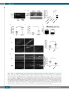
Figure 1. Migfilin-/- mice display impaired hemostatic and thrombotic functions. (A) Migfilin mRNA expression in wild-type (WT) platelets and exon 7 (179 bp) deleted migfilin-/- mice platelets. (B) Analysis of migfilin protein expression by Western blotting in heart tissues and platelets from WT and migfilin-/- mice. (C) Quantitative real- time PCR results analysing the expression of migfilin in platelets and heart tissues. Data are represented as a ratio relative to an internal control (β-actin) (mean ± standard error of the mean [SEM], n=3), ***P<0.001, Student t test. (D) Bleeding times for WT (●) and migfilin-/- mice (▲). Means are indicated by horizontal lines. **P<0.01, evaluated with 2-tailed Mann-Whitney U tests. (E) Weight of blood loss from WT (●) and migfilin-/- mice (▲) during bleeding time. Results are expressed as the mean ± SEM (n = 9). *P<0.05, evaluated with 2-tailed Mann-Whitney U tests. (F) Percentages of WT and migfilin-/- mouse bleeding times exceeded 15 minutes (min) (□) or were within 15 min (■). Results were obtained from 21 WT and 22 migfilin-/- mice. (G) Representative images of thrombi in FeCl3-injured mesenteric arte- rioles at indicated post-injury time points in WT (upper row) and migfilin-/- mice (lower row). a, arteriole; v: venule. Scale bars, 100 mm (left panel). Occlusion times of FeCl3-induced thrombosis in arterioles of WT (n=16) and migfilin-/- mice (n=16). Means are indicated by horizontal lines. **P<0.01, 2-tailed Mann-Whitney test (right panel). (H) Adhesion of platelets from WT and migfilin-/- mice on collagen. Mepacrine-labeled whole blood from WT and migfilin-/- mice was perfused over collagen- coated bioflux plates at a shear rate of 40 dynes/cm2 for 5 min. Original magnification 10x. Scale bar, 100 mm (left panel). Area coverage of platelets from WT and migfilin-/- mice (n=10 for both groups) after 5 min perfusion over a collagen surface, **P<0.01, 2-tailed Mann-Whitney test (right panel).
2610
haematologica | 2020; 105(11)
