
Page 75 - 2020_09-Haematologica-web
P. 75
Impaired microRNA processing in RA neutrophils
A
C
B
D
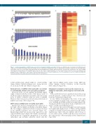
Figure 1. Global downregulation of miRNA expression profile in rheumatoid arthritis neutrophils. (A) Forty-one miRNA altered in neutrophils from PB-RA patients compared to neutrophils from PB-HD using a fold change cut-off >5. (B) Thirty-four miRNA more reduced in RA synovial neutrophils compared to its paired PB sample (fold change cut-off >2). (C) Heat-map of the differentially expressed miRNA profile in PB-HD, PB-RA and SF-RA. (D) Functional classification of the altered miRNA using Ingenuity Pathway Analysis (IPA, QIAGEN Redwood City, CA, USA; https://analysis.ingenuity.com). The analysis included only the functions and pathways with average IPA score >2 indicated as –log (P-value). Threshold bar indicates the cut-off point of significance (P>0.05), using Fisher’s exact test. miRNA: microRNA; RA: rheumatoid arthritis; PB: peripheral blood; HD: healthy donor; SF: synovial fluid.
to HD and PB paired samples (MIP-1 /1 , CCL5, CD40L, C5/5a, CXCL1, CXCL12, ICAM-1, IL-1 , IL-1ra, IL-8, IL- 13, IL-16, IL-18, MIF and TREM-1) (Figure 3D).
Reduced levels of miRNA in RA neutrophils are related to autoimmunity, clinical and serological parameters
Decreased levels of both, miRNA and DICER signifi- cantly correlated with the activity of the disease, levels of ACPA and clinical inflammatory markers. Elevated serum levels of TNF-a correlated with low levels of DICER. However, there was not association between the levels of miRNA and serum TNF-a (Figure 4A).
ACPA reduces miRNA levels in healthy neutrophils
Enriched IgG-ACPA downregulated the expression of the eight selected miRNA in healthy neutrophils (Figure 4B). Accordingly, a significant reduction of genes involved in the miRNA biogenesis was observed (Figure 4C). Enriched IgG-ACPA also increased the expression of the selected mRNA targets (Figure 4D). Finally, enriched IgG- ACPA promoted a significant upregulation of secondary chemokines and cytokines indirectly related with the
eight selected mRNA targets (CCL1, CCL2, MIP-1a/β, CCL5, CD40L, CXCL1, CXCL12, G-CSF, GM-CSF, IFN-g and IL-8) (Figure 4E).
Inflammatory mediators decrease the expression of miRNA in neutrophils, which might be restored by IFX or TCZ
TNF-a and IL-6 levels were significant elevated in serum from RA patients; a further increase was observed in SF from those RA patients (Figure 5A).
In vitro, TNF-a downregulated the levels of the eight selected miRNA alongside with a decrease in the expres- sion of DICER and AGO-2 in HD neutrophils (Figure 5B). Treatment with IL-6 reduced the levels of miR-126, let-7b, miR-17, AGO1 and AGO2 (Figure 5C).
We observed that fresh neutrophils from RA patients had significantly higher levels of TNF-a and IL-6 mRNA compared to freshly isolated neutrophils from HD (Online Supplementary Figure S5A). In addition, after 6 hours of in vitro culture, levels of TNF-a and IL-6 were elevated in the culture media of RA neutrophils (Online Supplementary Figure S5B).
haematologica | 2020; 105(9)
2253
