
Page 74 - 2020_09-Haematologica-web
P. 74
I.A. de la Rosa et al.
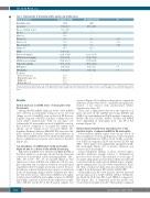
Table 1. Clinical details of rheumatoid arthritis patients and healthy donors.
Clinical parameters
Woman/Men (n/n)
Age (years)
Disease evolution (years) RF+ (%)
ACPA+ (%)
Obesity (%)
Diabetes (%) Hypertension (%) Smoker (%)
DAS28
Cholesterol (mg/mL) HDL-Cholesterol (mg/mL) LDL-Cholesterol (mg/mL) Triglycerides (mg/mL) ESR (mm/h)
CRP (mg/mL)
Treatments Corticosteroids (%) Hydroxychloroquine (%) NSAIDs (%) Methotrexate (%)
RA patients (n=40)
26/14
55.54 ± 4.03 9.20 ± 2.23 57.50 65.00 8.00 10.00 12.50 15.00 3.75 ± 0.20 202.15 ± 12.65 43.16 ± 2.68 136.81 ± 11.01 99.50 ± 10.16 15.75 ± 2.89 10.23 ± 2.73
50.00 45.00 80.00 70.00
HD controls (n=40) P 22/18
0.035 0.033 0.048
48.50 ± 8.45
-
-
-
3.00
5.00
6.00 12.00
- 200.20 ± 15.60 53.63 ± 12.75 115.75 ±17.40 88.50 ± 25.39
4.65 ± 1.20 0.01 1.55 ± 0.22 0.03
- - - -
Values are mean ± standard deviation (SD). HDL: high density lipoprotein; LDL: low density lipoprotein; DAS: disease activity score; ACPA: antibodies to citrullinated protein anti- gens; RA: rheumatoid arthritis; HD: healthy donor; RF: rheumatoid factor; ESR: erythrocyte sedimentation rate; CRP: C reactive protein; NSAIDS: non-steroideal anti-inflammatory drugs.
Results
Global decrease in miRNA levels of neutrophils from RA patients
Among the 800 miRNA analyzed, levels of 133 miRNA were detected in neutrophils. Using an above two-fold change cut-off, 94 miRNA were reduced in PB-RA neu- trophils compared to PB-HD, and three of them were ele- vated (Online Supplementary Table S1 and Figure 1A). Additionally, SF neutrophils showed 34 miRNA even fur- ther reduced compared to its paired PB sample (fold change cut-off change above two-fold) (Figure 1B). Ingenuity Pathway Analysis (QIAGEN IPA) uncovered the main enriched biological functions and pathways in which these miRNA are involved, which include immune disease, inflammatory response and connective disorders (Figure 1D).
Low abundance of miRNA levels in RA neutrophils might be due to a defect in the miRNA processing
Eight altered miRNA were identified by QIAGEN IPA as the main regulators of proteins involved in the abnormal activation of neutrophils in RA, including miRNA -126, - 148a, -29c, let-7b, -30c, -17, -21 and 223 (Figure 2). The expression of these miRNA was validated in all the sam- ples separately and a technical validation was performed separately in the 10 samples previously used for the pool. In addition, a clinical validation was carried out separately in the 30 remaining samples (Online Supplementary Figure S2). The levels of most of the selected miRNA were signif- icantly reduced in PB-RA neutrophils compared to PB-HD neutrophils. A greater reduction in the expression of miR- 148a, miR-29c and let-7b in the SF paired samples was
observed (Figure 3A). In addition, there was not significant difference in the reduced levels of miRNA among patients treated or not treated with methotrexate (Online Supplementary Figure S3).
There was a significant reduction in the expression of genes involved in the miRNA processing (DICER and AGO-1) in neutrophils from PB-RA patients compared to PB-HD. Of note, DICER, AGO-1, AGO-2 and XPO-5 were diminished in neutrophils from the SF of RA patients (Figure 3B).
Bioinformatic identification and expression of the putative targets of reduced miRNA in RA neutrophils
Seven putative mRNA targets were chosen based on their recognized role in the pathogenesis of RA, being key factors in inflammation (TNF-a, IL-1β, IL-6R), cell adhe- sion (VEGF-A), migration (IL-8) and survival (STAT3 and AKT). These targets were significantly upregulated in PB- RA neutrophils (Figure 3C). A greater alteration was observed in SF neutrophils.
Using enrichment analysis of those selected targets, enriched pathways mainly related to inflammatory processes were revealed. This included a broad range of secondary chemokines and cytokines which are indirectly connected with the eight selected mRNA targets, amplify- ing the inflammatory cascade (Online Supplementary Figure S4). Thus, a human cytokine array was performed in neu- trophils from RA patients (PB and SF) and HD (PB).
Neutrophils from PB of RA patients showed increased protein expression of CCL5, CD40L, CXCL1, CXCL2, IL- 1ra, IL-16, IL-18, IL-32a, PAI-1 and TREM-1 compared to HD (Figure 3D). A differential proteome profile was observed in neutrophils from SF of RA patients compared
2252
haematologica | 2020; 105(9)
