
Page 42 - 2020_09-Haematologica-web
P. 42
P. Bianchi and E. Fermo
The number of known pathogenic variants is continu- ously increasing. In a recent inventory, Canu et al.33 report- ed 260 mutations in the PKLR gene; the Human Genome Mutation Database (HGMD) reports 290 pathogenic vari- ants (update March 2020); a detailed inventory of PKLR variants is also available in the PKLR Leiden Open Variation Database (https://databases.lovd.nl/shared/genes/ PKLR), including a more specific data collection (e.g., con- gress presentations and unpublished results).
The HGMD does not yet include the results obtained in a single analysis of 257 patients with PK deficiency enrolled in the Pyruvate Kinase Deficiency Natural History Study (PKD NHS), a multicenter, international study; 127 different pathogenic variants were detected, comprising 84 missense and 43 non-missense variants (including 20 stop-gain variants, 11 affecting splicing, 5 large deletions, 4 in-frame indels, and 3 promoter vari- ants).34,35 A similar distribution is observed by stratifying variants reported by the HGMD according to the type of mutations (Figure 2).
A list of the more commonly detected mutations and variants with geographical distribution and ethnic back- ground is reported in Table 2; similar information for the rarer/unique variants is available in mutation databases. Molecular analysis of the PKLR gene by Sanger sequenc- ing usually covers the entire coding region, flanking intronic sequences and the erythroid-specific promoter. NGS analysis allows more extensive sequencing than the Sanger method (generally including entire coding and intronic flanking regions, promoter, 3’ upstream, and 5’ downstream regions) and can give information on the presence of large indels. Other techniques (e.g., multiplex ligation-dependent probe amplification or assays of copy number variations, comparative genomic hybridization arrays or digital polymerase chain reaction) can also be used to this latter purpose. Variants are usually reported in the literature using the RPK cDNA sequence of the PKLR gene, with the A of the initiation ATG being assigned number +1 (Transcript refseq ID NM_000298.5). American College of Medical Genetics and Genomics (ACMG) guidelines should be followed to interpret and assess sequence variants.42
Promoter and enhancer variants
Only a few pathogenic variants have been identified in the promoter region, mostly clustering at two functionally important sequences, such as the consensus binding motif for GATA-1 at nucleotides c.-69 to -74,43-45 and a regulatory element (PKR-RE1) whose core CTCTG extends from nucleotides c.-87 to -83.46,47 The variant c.-72A>G, located in the GATA-1 motif, was found to be associated with low mRNA expression, and to be responsible for severe ane- mia when present in the homozygous state.15,45 Other vari- ants have been reported with uncertain pathogenic signif- icance: the variant c.-109C>T described by Pissard et al.,48 while not directly modifying any known binding site for a transacting factor, was found to be located within a region displaying basic promoter activity, very close to the region described as an erythroid enhancer49 (Figure 3).
At the moment a clear disease-causing association of variants located in the enhancer region is not well estab- lished. Some reported variants in this regions, such as c.- 148C>T,49 or deletion reported at nucleotides c.-249delA or c.-248delT, do not seem to affect the expression of the gene, thus are considered non-pathogenic.46
Coding region variants
The large majority of pathogenic variants are located in the coding region. Mutations are distributed throughout the PKLR gene and affect all exons (Figure 4). Most of them (about 66%) are missense mutations (Figure 2). Not every mutation detected by DNA sequencing can be immediately classified as a disease-causing variant, and should be considered ‘variants of unknown clinical signif- icance’ until their pathogenic nature is confirmed by func- tional analysis such as PK enzymatic assays, western blot- ting, reverse transcriptase polymerase chain reaction analysis, or gene reporter assays.50 This is especially important when patients’ samples are not accompanied by complete clinical and laboratory information.
Most variants in PK deficiency affect residues critical to the structure and/or function of the enzyme. However, it is not possible to establish a direct relationship between the severity of a pathogenic variant and its position; most patients in fact are compound heterozygous for two
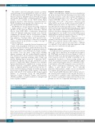
Table 1. Recent studies performed by next-generation sequencing technologies in patients with hemolytic anemias.
Reference Method N. of genes N. of cases studied analyzed with CHA
15 t-NGS 35 36
28 t-NGS 55 43 29 WES n.a. 4 30 t-NGS 76 21
b 27 t-NGS 76 21
25 t-NGS 34 and 71 74c 23 t-NGS 33 57
Number of genes included in the panel, number of cases analyzed in each study and cases diagnosed with pyruvate kinase deficiency are shown. Next-generation sequencing analysis allowed modification of a previous diagnosis; the number and the type of mismatched diagnosis is reported in the last column. aAll transfusion-dependent patients. bNo diagnosis despite extensive laboratory investigations. cSuspected diagnosis of congenital dyserythropoietic anemia. CHA: chronic hemolytic anemias; PKD: pyruvate kinase defi- ciency; t-NGS: targeted next-generation sequencing; WES: whole-exome sequencing; n.a.: not available; CDA: congenital dyserythropoietic anemia; DBA: Diamond-Blackfan ane- mia.
a
PKD diagnosis New diagnosis and number and type of mismatched diagnoses
2 2newPKD
8 8newPKD
4 4newPKD
6 3newPKD 2 CDA→ PKD 1 DBA→PKD
6 4newPKD 2 CDA→PKD
7 7 CDA→PKD 3 2newPKD
1 CDA→PKD
2220
haematologica | 2020; 105(9)
